组成图示
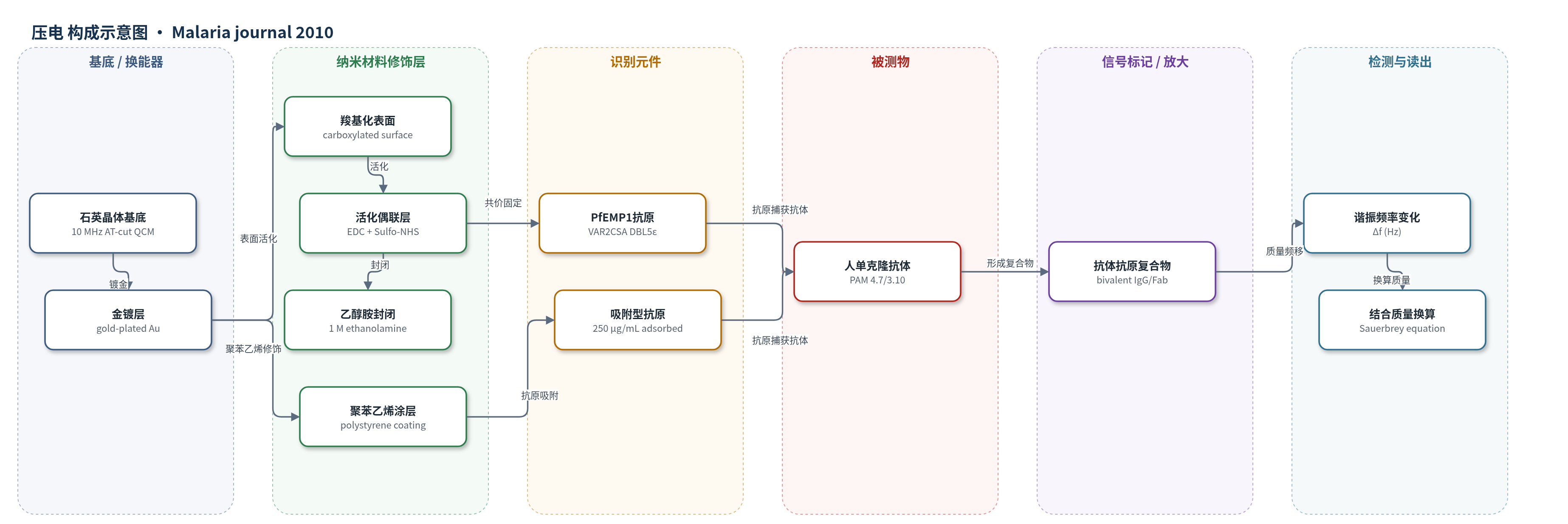
点击图片查看大图 · 依据论文自动绘制
传感器类型
压电(QCM)生物传感器
检测对象
人抗 PfEMP1 单克隆抗体(human anti-PfEMP1 monoclonal antibodies, PAM 4.7、PAM 3.10 及其 Fab 片段),样品基质为 PBS pH 7.4 流动缓冲液(聚苯乙烯体系含 1 g/L BSA)。
检测原理
该传感器以压电石英晶体为换能器:晶体表面质量增加时,谐振频率下降,按 Sauerbrey 方程将频率变化换算为结合质量。先将重组 VAR2CSA PfEMP1 DBL5ε 抗原通过 EDC/Sulfo-NHS 共价偶联到羧基化金表面,或吸附到聚苯乙烯表面,形成识别层;流动相中的人单克隆抗体或 Fab 片段与固定抗原结合,使表面质量增加,频率下降;解离时质量减少,频率回升。抗体浓度升高时,结合速率和平衡结合量增加。二价 IgG 可交联相邻抗原表位,产生亲合力放大,使表观解离变慢;Fab 单价结合消除交联,可测单表位解离。整个检测无标记、实时进行,不依赖酶催化或荧光放大。
检测灵敏度
未报告 LOD、线性范围、灵敏度斜率或相关系数。
效应效果
QCM 在流动条件下无标记实时监测抗体结合,空白基线稳定,重复注入可评估芯片损伤。PAM 4.7 对固定 VAR2CSA DBL5ε 的 KD 为 2.91×10^-8 M;PAM 3.10 在高密度表面 kon 为 1.72×10^5 M^-1s^-1,低密度表面为 4.2×10^6 M^-1s^-1,koff 低于仪器可测限(<10^-6 s^-1),估算 KD<2.38×10^-13 M。PAM 3.10 Fab 单价结合 koff 为 4.04×10^-5 s^-1,说明整抗体极低解离主要来自二价亲合力。AFM 测得 knob 直径约 120 nm、高约 24 nm、表面积约 13,000 nm²,估算最多约 110 个 VAR2CSA、密度约 8,000 个/μm²。作者认为该体系可指导抗黏附疫苗诱导高亲合力、低解离抗体。
传感器的构成
- 基底/换能器:10 MHz AT-cut 石英晶体(gold-plated quartz crystal),提供压电谐振与质量传感
- 金镀层:Au 表面,作为蛋白固定与界面修饰基底
- 表面化学修饰:羧基化表面(carboxylated surface)或聚苯乙烯涂层(polystyrene coating),用于共价偶联或吸附抗原
- 活化偶联层:EDC 与 Sulfo-NHS 活化羧基,形成氨基反应性酯,用于固定 VAR2CSA DBL5ε 抗原
- 识别元件:重组 VAR2CSA PfEMP1 DBL5ε 抗原(40.1 kDa,339 aa)固定于芯片表面,捕获流动相中抗体
- 封闭剂:1 M 乙醇胺(ethanolamine, pH 8.0)封闭剩余活化位点;聚苯乙烯体系流动缓冲液含 1 g/L BSA 降低非特异结合
- 信号读出:QCM 谐振频率变化(Hz),经 Sauerbrey 方程换算结合质量,无标记检测抗体结合/解离
中文摘要
背景:感染疟疾的人类会对疟原虫输出至红细胞膜的 PfEMP1 黏附抗原产生保护性抗体反应,但抗体-受体结合动力学及 PfEMP1 膜拓扑如何影响抗体结合与解离仍不清楚。方法:采用石英晶体微天平(QCM)生物传感器,在流动条件下实时测量 VAR2CSA PfEMP1 与人单克隆抗体的结合和解离动力学;用免疫荧光显微镜观察活感染红细胞表面抗体介导黏附;用原子力显微镜(AFM)获得膜 knob 高分辨图像,估算 knob 表面积并建模 VAR2CSA 堆积密度。结果:高亲合力相互作用时,抗体从 VAR2CSA PfEMP1 解离极慢;高亲合力结合需要抗体二价交联其可桥接距离内的表位。knob 表面积与 PfEMP1 堆积密度计算表明,高亲合力交联受 knob 结构和 PfEMP1 大分子尺寸限制。结论:强结合 VAR2CSA PfEMP1 需要高亲合力,但 knob 结构会抑制抗原间交联,因许多表位位于抗体 15–18 nm 扫描半径之外;PfEMP1 大尺寸也限制 knob 内交联。提示疫苗应诱导强结合、高亲合力抗体,其解离速率常数比结合速率常数更重要。
英文摘要
BACKGROUND: Infected humans make protective antibody responses to the PfEMP1 adhesion antigens exported by Plasmodium falciparum parasites to the erythrocyte membrane, but little is known about the kinetics of this antibody-receptor binding reaction or how the topology of PfEMP1 on the parasitized erythrocyte membrane influences antibody association with, and dissociation from, its antigenic target.
METHODS: A Quartz Crystal Microbalance biosensor was used to measure the association and dissociation kinetics of VAR2CSA PfEMP1 binding to human monoclonal antibodies. Immuno-fluorescence microscopy was used to visualize antibody-mediated adhesion between the surfaces of live infected erythrocytes and atomic force microscopy was used to obtain higher resolution images of the membrane knobs on the infected erythrocyte to estimate knob surface areas and model VAR2CSA packing density on the knob.
RESULTS: Kinetic analysis indicates that antibody dissociation from the VAR2CSA PfEMP1 antigen is extremely slow when there is a high avidity interaction. High avidity binding to PfEMP1 antigens on the surface of P. falciparum-infected erythrocytes in turn requires bivalent cross-linking of epitopes positioned within the distance that can be bridged by antibody. Calculations of the surface area of the knobs and the possible densities of PfEMP1 packing on the knobs indicate that high-avidity cross-linking antibody reactions are constrained by the architecture of the knobs and the large size of PfEMP1 molecules.
CONCLUSIONS: High avidity is required to achieve the strongest binding to VAR2CSA PfEMP1, but the structures that display PfEMP1 also tend to inhibit cross-linking between PfEMP1 antigens, by holding many binding epitopes at distances beyond the 15-18 nm sweep radius of an antibody. The large size of PfEMP1 will also constrain intra-knob cross-linking interactions. This analysis indicates that effective vaccines targeting the parasite's vulnerable adhesion receptors should primarily induce strongly adhering, high avidity antibodies whose association rate constant is less important than their dissociation rate constant.