组成图示
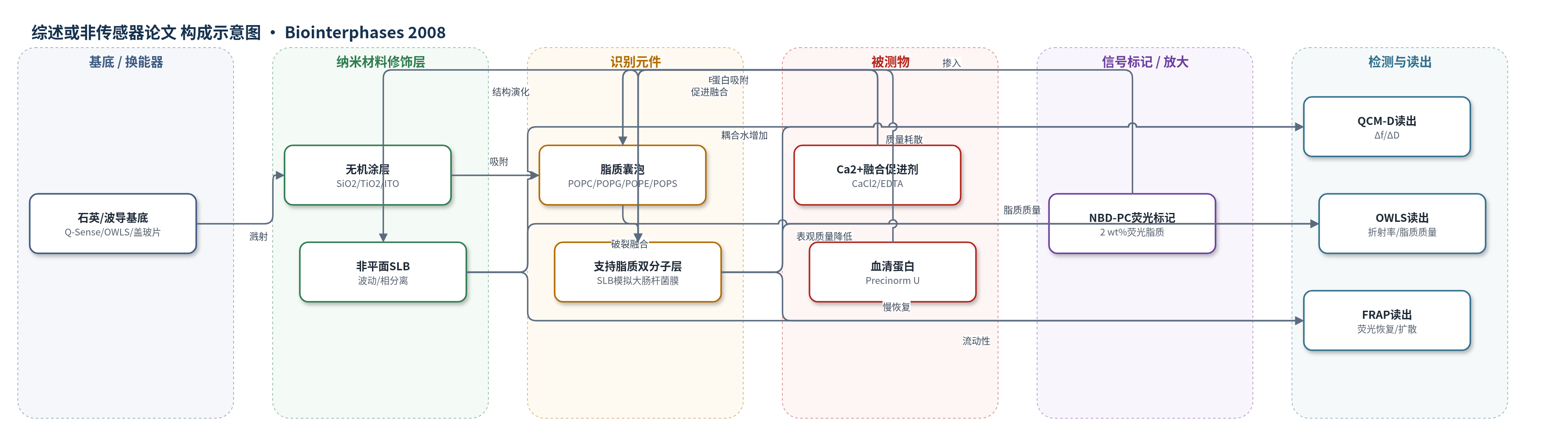
点击图片查看大图 · 依据论文自动绘制
传感器类型
综述或非传感器论文
检测对象
无明确检测分析物;研究膜前驱体:大肠杆菌总脂质提取物(E. coli total lipid extract)、POPC/POPG/POPE/POPS 脂质体;样品基质:10 mM HEPES/150 mM NaCl/CaCl2 缓冲液、血清(Precinorm U)
检测原理
本文并非以单一分析物浓度产生传感信号,而是研究仿生膜在传感器基底上的形成机制。带负电的脂质囊泡在 SiO2、TiO2 或 ITO 表面通过静电作用吸附;Ca2+ 在界面富集并中和/桥接阴离子脂质头基,降低囊泡-表面排斥,促进囊泡变形、破裂和融合,形成支持脂质双分子层(SLB)。QCM-D 通过压电晶体频率下降和耗散变化反映吸附脂质及耦合水的质量与膜柔顺性;OWLS 通过倏逝场有效折射率变化计算脂质干质量;FRAP 利用 NBD-PC 荧光漂白后恢复反映膜横向连通性和扩散。Ca2+ 浓度过低时仅囊泡吸附,适中时形成 SLB,过高时引起聚集或非平面波动,使 QCM-D 耦合水增加、OWLS 表观质量下降、FRAP 恢复变慢。
检测灵敏度
原文未报告 LOD、线性范围、灵敏度斜率或相关系数。
效应效果
作者在 SiO2、TiO2 和 ITO 上筛选 CaCl2 条件,测量重复两次以上。SLB 典型 QCM-D 响应为 Δf 约 −24 至 −27 Hz(约 450 ng/cm2)、ΔD 约 0.3×10⁻⁶;OWLS 脂质质量接近 310 ng/cm2,每脂质面积约 75 Å2;FRAP 恢复约 90±10%,扩散系数约 5×10⁻⁹ cm2/s,部分 SLB 约 30%,囊泡无恢复。E. coli 总脂质在 TiO2 上 0.2–2 mM CaCl2 可形成 SLB 或非平面 SLB,血清吸附被抑制,部分 SLB 上血清吸附可超过 150 ng/cm2。ITO 上未形成完整细菌模拟 SLB,但 POPC:POPS 可形成紧密 SLB。作者认为可用于抗生素与细菌膜相互作用研究。
传感器的构成
- 基底/换能器:5 MHz AT-cut 石英晶体(Q-Sense)、OWLS 波导(MicroVacuum)或盖玻片,提供压电、光学波导或荧光成像基底
- 无机涂层:50 nm SiO2、12 nm TiO2 或 50 nm ITO(In2O3 90%/SnO2 10%),磁控溅射形成带表面电荷的脂质吸附界面
- 脂质囊泡:POPC、POPC:POPS、POPC:POPG、POPE:POPG 或 E. coli total lipid extract 脂质体,50 μg/mL,作为支持膜前驱体
- 支持脂质双分子层:由囊泡破裂融合形成的 SLB,模拟大肠杆菌膜组成,远端叶暴露于缓冲液
- 荧光标记:NBD-PC(2 wt%)掺入脂质体,用于 FRAP 监测膜流动性与横向扩散
- 缓冲/融合促进剂:10 mM HEPES、150 mM NaCl,CaCl2 0–20 mM 或 2 mM EDTA,调节静电屏蔽与 Ca2+ 融合
- 读出层:QCM-D(Δf/ΔD)、OWLS(有效折射率/脂质质量)和 FRAP(荧光恢复),表征膜形成、质量与流动性
中文摘要
近年来,表面支持脂质双分子层(SLB)通过脂质体在固体表面破裂融合形成,已成为研究细胞膜生物物理与生化相互作用的重要模型。本文报道了强负电荷支持脂质膜的制备与表征,其脂质组成系统模拟大肠杆菌(E. coli)膜,并在三种常用生物传感器材料二氧化硅(SiO2)、二氧化钛(TiO2)和氧化铟锡(ITO)上进行研究。作者通过调节 Ca2+ 浓度平衡脂质囊泡电荷与界面静电作用,优化囊泡吸附、破裂和融合过程,获得脂质囊泡-表面相互作用的新认识。吸附过程采用带耗散监测的石英晶体微天平(QCM-D)、光波导光模光谱(OWLS)和光漂白后荧光恢复(FRAP)进行互补表征,能够识别平面与非平面支持脂质膜的形成。该工作为复杂脂质组成仿生膜涂层在药物筛选、动态生物界面、人工鼻及膜活性抗生素研究中的应用提供了基础。
英文摘要
In recent years, a large effort has been spent on advancing the understanding of how surface-supported membranes are formed through vesicle fusion. The aim is to find simple model systems for investigating biophysical and biochemical interactions between constituents of cell membranes and, for example, drugs and toxins altering membrane function. Designing and controlling the self-assembly of model membranes onto sensor substrates thus constitutes an important field of research, enabling applications in, e.g., drug screening, dynamic biointerfaces, artificial noses, and research on membrane-active antibiotics. The authors have developed and investigated the formation of strongly negatively charged supported lipid membranes which systematically mimic bacterial membrane composition on three important biosensor materials: SiO(2), TiO(2), and indium tin oxide. By tuning the electrostatic interaction through balancing the lipid vesicle charge with the ionic strength of Ca(2+) as a fusion promoter, the authors have optimized the self-assembly and obtained new insights into the details of lipid vesicle-surface interaction. The results will be useful for future development and application of specialized lipid membrane surface coatings prepared from complex lipid compositions. The adsorption processes were characterized by a quartz crystal microbalance with dissipation monitoring, optical waveguide lightmode spectroscopy, and fluorescence recovery after photobleaching, which allowed the determination of formation also of nonplanar supported lipid membranes.