组成图示
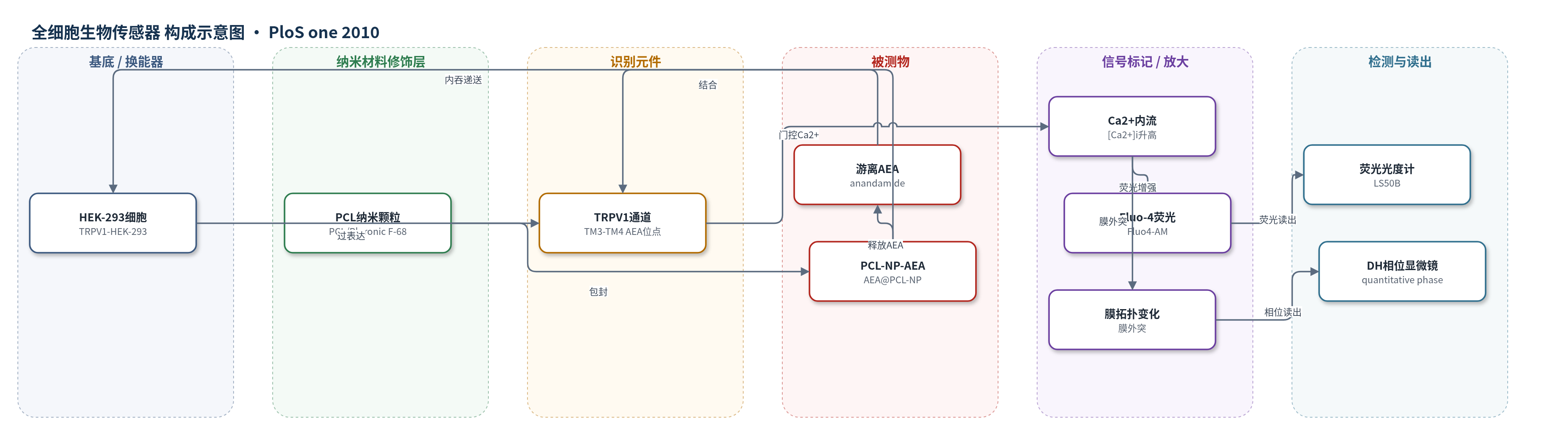
点击图片查看大图 · 依据论文自动绘制
传感器类型
全细胞生物传感器
检测对象
花生四烯酸乙醇胺(anandamide, AEA;游离AEA及PCL-NP-AEA递送形式),样品基质为完整TRPV1-HEK-293细胞培养液(Tyrode’s solution/EMEM)
检测原理
AEA进入细胞后结合TRPV1通道TM3–TM4区胞质位点,使通道门控开放,胞外Ca2+内流。Fluo-4探针与Ca2+结合后荧光增强,荧光强度随AEA浓度及摄取量增加而升高;数字全息相位显微镜则通过Ca2+内流引发的细胞表面膜外突/相位变化提供无标记读出。PCL-NPs将AEA包封并内吞进入细胞,在胞质释放AEA后同样激活TRPV1,从而绕过质膜转运蛋白。比较游离AEA与PCL-NP-AEA对摄取抑制剂的敏感性,可区分膜转运体介导的摄取与胞内递送途径。
检测灵敏度
未报告LOD、线性范围、灵敏度斜率或R^2。
效应效果
在TRPV1-HEK-293细胞中,1 μM AEA使[Ca2+]i达ionomycin效应的72.0±61%,capsaicin为76±8%,仅被I-RTX拮抗,对BSA、OMDM-1、MbCD、OA、FABP4抑制剂不敏感。PCL-NP-AEA药效/药代与游离AEA相近,100 nM效应10天稳定,游离AEA第10天降低50.5±4.3%;PCL-NPs包封率96.05±1.77%。PCL-NP-AEA对OMDM-1、AM1172、BSA敏感性显著降低,支持AEA转运体存在。
传感器的构成
- 细胞基底:HEK-293细胞(TRPV1-HEK-293,稳定过表达人重组TRPV1),提供完整质膜与胞质环境
- 识别/换能元件:TRPV1通道(TM3–TM4胞质AEA结合位点),结合AEA后门控Ca2+内流
- 信号标记物:Fluo-4 AM(Fluo4-AM)钙离子荧光探针,Ca2+升高引起荧光增强
- 纳米递送层:PCL-NPs(poly-ε-caprolactone nanoparticles,Pluronic F-68包覆),负载AEA或Rhodamine-123并介导胞内递送
- 荧光读出:Perkin-Elmer LS50B荧光分光光度计(λEx 488 nm/λEm 516 nm),记录[Ca2+]i
- 相位读出:数字全息定量相位显微镜(DH quantitative phase microscopy),记录Ca2+内流后细胞表面拓扑变化
中文摘要
本文报道利用TRPV1通道的胞质花生四烯酸乙醇胺(AEA)结合位点作为生物传感器,检测AEA进入完整TRPV1过表达HEK-293细胞,并结合纳米技术研究其膜转运。作者采用Fluo-4荧光法测量胞内Ca2+升高,以及数字全息定量相位显微镜观察Ca2+内流引起的细胞表面拓扑变化,以表征TRPV1激活。将AEA包封于聚己内酯纳米颗粒(PCL-NPs)中,可使其绕过可能参与摄取的膜蛋白进入细胞并激活TRPV1。若不存在特异性转运蛋白,则此前报道的AEA摄取抑制剂应对游离AEA和PCL-NP-AEA产生相同抑制;但结果显示,PCL-NP-AEA的细胞摄取对BSA、OMDM-1、AM1172、MbCD、油酸及FABP4抑制剂等的敏感性显著降低,而药效学与药代动力学仍与游离AEA相近。这些结果支持上述抑制剂对假想AEA转运体具有特异性,并间接支持AEA载体介导膜转运机制的存在。
英文摘要
BACKGROUND: Considerable efforts have been made to characterize the pathways regulating the extracellular levels of the endocannabinoid anandamide. However, none of such pathways has been so argued as the existence of a carrier-mediated transport of anandamide across the membrane. Apart from the lack of molecular evidence for such a carrier, the main reasons of this controversy lie in the methodologies currently used to study anandamide cellular uptake. Furthermore, the main evidence in favor of the existence of an "anandamide transporter" relies on synthetic inhibitors of this process, the selectivity of which has been questioned.
METHODOLOGY/PRINCIPAL FINDINGS: We used the cytosolic binding site for anandamide on TRPV1 channels as a biosensor to detect anandamide entry into cells, and exploited nanotechnologies to study anandamide membrane transport into intact TRPV1-overexpressing HEK-293 cells. Both fluorescence and digital holographic (DH) quantitative phase microscopy were used to study TRPV1 activation. Poly-epsilon-caprolactone nanoparticles (PCL-NPs) were used to incorporate anandamide, which could thus enter the cell and activate TRPV1 channels bypassing any possible specific protein(s) involved in the uptake process. We reasoned that in the absence of such protein(s), pharmacological tools previously shown to inhibit the "anandamide transporter" would affect in the same way the uptake of anandamide and PCL-NP-anandamide, and hence the activation of TRPV1. However, when masked into PCL-NPs, anandamide cellular uptake became much less sensitive to these agents, although it maintained the same pharmacokinetics and pharmacodynamics as that of "free" anandamide.
CONCLUSIONS: We found here that several agents previously reported to inhibit anandamide cellular uptake lose their efficacy when anandamide is prevented from interacting directly with plasma membrane proteins, thus arguing in favor of the specificity of such agents for the putative "anandamide transporter", and of the existence of such mechanism.