组成图示
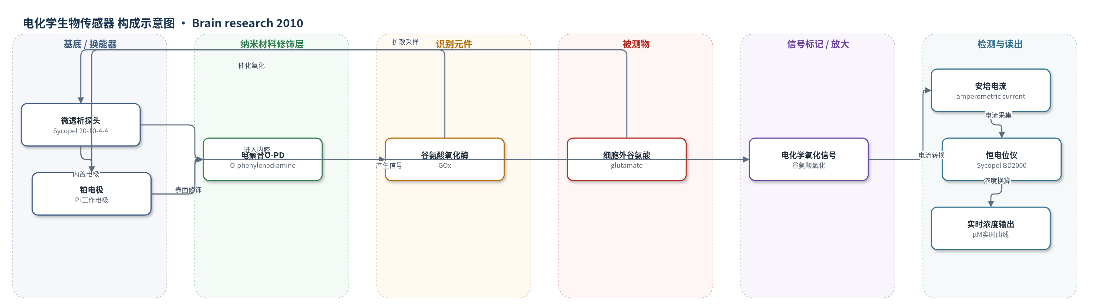
点击图片查看大图 · 依据论文自动绘制
传感器类型
电化学生物传感器
检测对象
细胞外谷氨酸(glutamate, Glu);样品基质:大鼠脑组织细胞外液(微透析液)
检测原理
该传感器基于微透析采样与酶促安培检测。大鼠脑细胞外液中的谷氨酸通过Sycopel微透析膜扩散进入探头内腔,内腔以0.5 μL/min灌注含谷氨酸氧化酶(GOx)的PBS。GOx催化谷氨酸氧化,在电聚合邻苯二胺(O-PD)修饰的铂电极(Pt)表面产生可检测的电化学信号。恒电位仪采集安培电流,电流大小与谷氨酸浓度成正比。标准谷氨酸溶液中传感器在50–450 μM呈线性响应,灵敏度为0.22 nA/μM,R^2=0.998。缺血发生后,细胞外谷氨酸释放增加,传感器电流随之升高,从而实现对脑内谷氨酸动态变化的实时监测。
检测灵敏度
线性范围: 50–450 μM;灵敏度: 0.22 nA/μM;R^2 = 0.998
效应效果
传感器在标准谷氨酸溶液中具有50–450 μM线性范围、0.22 nA/μM灵敏度和R^2=0.998。11VO模型中,闭塞后脑血流降至13.5±6.1%,缺血平台15.3±2.5 s内达到;谷氨酸自缺血后111.3±30.0 s升高。5 min组缺血期最大谷氨酸84.0±32.8 μM,10 min组140.0±24.3 μM;再灌注峰值95.0±30.7 μM和141.7±21.6 μM。72 h后,10 min组海马CA1存活细胞20.5±4.8%,5 min组79.2±6.7%;NeuN为正常组38.0±3.4%,C.caspase-3为缺血组7.0±1.3%。结果支持传感器用于脑缺血谷氨酸实时监测。
传感器的构成
- 微透析采样层:Sycopel General 20-10-4-4 微透析探头,选择性允许细胞外谷氨酸进入内腔
- 换能器电极:铂电极(Pt),作为安培检测工作电极
- 电化学修饰层:电聚合邻苯二胺(O-phenylenediamine, O-PD),在Pt电极表面形成修饰层
- 识别元件:谷氨酸氧化酶(glutamate oxidase, GOx),催化谷氨酸氧化
- 流动相:磷酸盐缓冲液(PBS),以0.5 μL/min灌注,维持酶活性并输送反应物
- 读出模块:Sycopel BD2000 恒电位仪,施加电位并采集安培电流
中文摘要
本研究旨在明确谷氨酸释放对大鼠十一血管闭塞(11VO)缺血模型中神经元细胞死亡的影响。采用250–350 g雄性Sprague–Dawley大鼠,建立5 min和10 min短暂闭塞的全脑缺血模型。手术过程中,利用微透析安培生物传感器实时测定细胞外谷氨酸浓度,并同步监测脑血流。缺血72 h后取脑,采用双免疫荧光检测神经元特异性核蛋白(NeuN)和裂解caspase-3(C.caspase-3)水平。结果显示,两种闭塞模型中脑血流百分比均显著下降;缺血开始后谷氨酸释放增加并持续升高,10 min组谷氨酸释放水平显著高于5 min组。与5 min组轻微脑损伤不同,10 min组谷氨酸升高导致海马区缺血性神经元死亡,伴C.caspase-3激活和NeuN表达抑制。研究提示,在11VO缺血模型中,谷氨酸释放增加可诱导凋亡性神经元死亡。
英文摘要
The aim of this study was to define the effects of glutamate release on cell death in an eleven vessel rat occlusion model. Male Sprague-Dawley rats (250-350g) were used for the 11 vessel occlusion ischemic model, which was induced by a 5- and 10-min transient occlusion. During the surgical procedure, the extracellular glutamate concentration was measured in real-time using a microdialysis amperometirc biosensor with cerebral blood flow. In order to confirm neuronal cell death, brains were removed 72h after ischemia for the detection of the neuron-specific nuclear protein and cleaved caspase-3 levels, using double-immunofluorescence. A significant decrease in % cerebral blood flow was observed in both the 5- and 10-min 11 vessel occlusion models, while an increase in glutamate release was detected after the onset of ischemia that continued to rise during the ischemic period. However, a significantly higher level of glutamate release was observed in the 10-min ischemia group compared to the 5-min group. Unlike the small amount of brain damage in the 5-min group, the increased glutamate levels in the 10-min group resulted in ischemic cell death in the hippocampal region with the activation of cleaved caspase-3 and the inhibition of neuron-specific nuclear protein expression. This study suggests that the increased level of glutamate release induces apoptotic cell death in the 11 vessel occlusion ischemic model.