组成图示
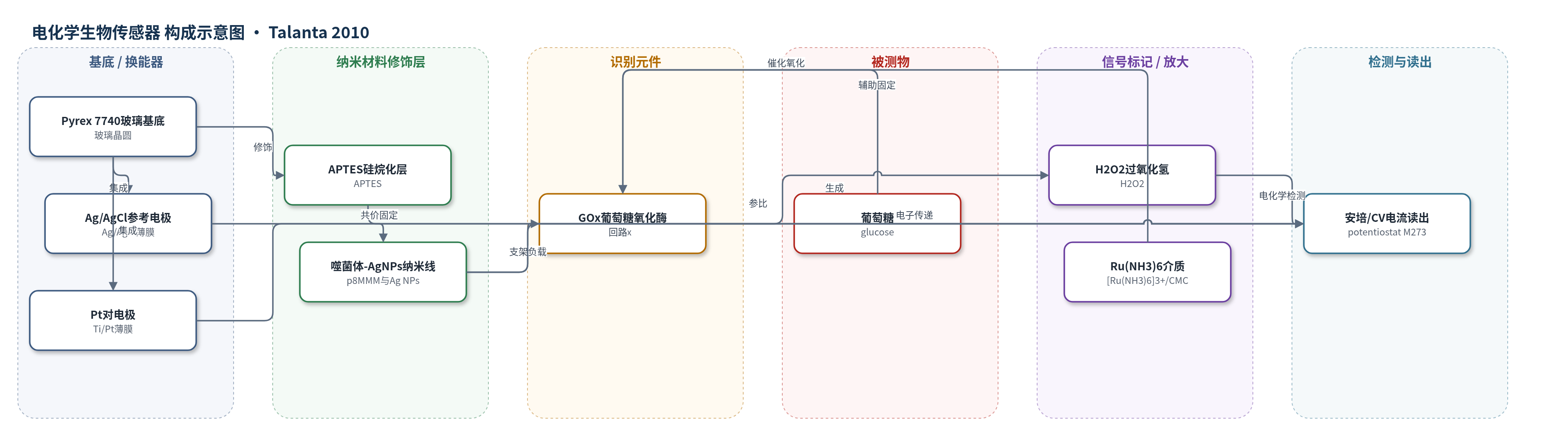
点击图片查看大图 · 依据论文自动绘制
传感器类型
电化学生物传感器
检测对象
葡萄糖(glucose);样品基质:磷酸盐缓冲液/搅拌缓冲液(pH 7.0/7.4)
检测原理
该传感器以GOx为催化识别元件,葡萄糖在溶解氧存在下被GOx催化氧化为葡萄糖酸并生成H2O2。p8MMM噬菌体表面展示的MMM肽通过甲硫氨酸介导的银结合与Ag纳米颗粒连接,形成生物纳米线网络;Ag纳米颗粒提供高导电通道,噬菌体网络增大工作电极有效面积并促进电子转移。GOx与[Ru(NH3)6]3+、CMC原位染色形成酶电极,H2O2在+0.3 V(vs. Ag/AgCl)下发生电化学反应,产生与葡萄糖浓度成正比的安培电流。Ag-p8MMM修饰使有效面积较Au纳米颗粒电极提高约1.85倍,从而增强灵敏度。
检测灵敏度
线性范围: 10^-7–10^-4 M;斜率: 19.97 μA cm^-2 mM^-1 (Ag-p8MMM)、10.21 μA cm^-2 mM^-1 (p8MMM)
效应效果
Ag-p8MMM生物纳米线电极在10^-7–10^-4 M葡萄糖范围内呈线性响应,斜率为19.97 μA cm^-2 mM^-1,高于p8MMM电极的10.21 μA cm^-2 mM^-1;CV峰电流和峰电位为689 μA/cm^2和280 mV。循环伏安有效面积较Au纳米颗粒电极提高1.85倍,峰-峰电位分离降低,表明电子转移能力增强;在-0.4至+0.8 V范围内连续100次CV循环后稳定性良好。pH 6.0–9.0中6.5–8.5响应最佳,温度20–50 C中25–35 C响应相近,超过35 C因GOx部分失活而下降。作者认为该平台可用于分析系统、无标记检测和生物传感器。
传感器的构成
- 基底:Pyrex 7740玻璃晶圆,经NaOH、丙酮、乙醇和去离子水清洗,承载三电极。
- 参考电极:Ag/AgCl薄膜,提供稳定参比电位。
- 对电极:Ti/Pt薄膜(Ti约200 Å、Pt约2500 Å),完成电化学回路。
- 工作电极修饰层:3-aminopropyltriethoxysilane (APTES)硅烷化层,提供氨基共价结合位点。
- 生物纳米线层:fd-tet p8MMM丝状噬菌体(pVIII展示MMM肽)与Ag纳米颗粒(6–7 nm)复合网络,作为导电支架并增大有效面积。
- 识别/催化层:葡萄糖氧化酶(GOx),催化葡萄糖氧化生成H2O2。
- 酶电极介质:GOx、[Ru(NH3)6]3+和0.1 wt%羧甲基纤维素(CMC)原位染色层,固定酶并辅助电子传递。
- 工作区限定:聚二甲基硅氧烷(PDMS)疏水环,限定反应区。
中文摘要
本文提出一种以基因工程改造丝状噬菌体与纳米颗粒构成的生物纳米线作为工作电极活性区,用于检测电化学反应的新方法。在生物传感器中,工作电极纳米材料的选择对灵敏度至关重要。作者将表面主要衣壳蛋白pVIII展示MMM肽的fd-tet p8MMM丝状噬菌体通过化学结合固定于电化学传感器活性区,形成由p8MMM噬菌体和银纳米颗粒组成的生物纳米线网络,用于特定分子的灵敏快速检测。以葡萄糖电化学检测为例,该生物纳米线传感器在10^-7–10^-4 M葡萄糖浓度范围内产生足够高的电流响应;循环伏安峰电流和峰电位分别为689 μA/cm^2和280 mV。该丝状纳米噬菌体电极在不同pH和温度下表现出高灵敏度和良好稳定性,有望用于分析系统、无标记检测和生物传感器。
英文摘要
This paper proposes a new approach to detect an electrochemical reaction using a working area consisting of bio-nanowires from genetically modified filamentous phages and nanoparticles. Use of the nanomaterials on the working electrode is a vital consideration in biological sensor development, because the biosensor sensitivity heavily depends on the material used. Here we use that fd-tet p8MMM filamentous phages displaying the MMM peptide on the major coat protein pVIII (designated p8MMM phages) were immobilized on the active area of an electrochemical sensor through chemical binding. The bio-nanowires composed of p8MMM phages and silver nanoparticles facilitated sensitive, rapid detection of particular molecules. We performed the experiment for observing electrochemical glucose detection to estimate the possibility of using one or other characterized-biological sensor. The current response of the bio-nanowire sensor reached sufficiently high signals at various glucose concentrations (10(-7) to 10(-4)M). The cyclic voltammetry peak current I(p) and peak potential E(p) were 689microA/cm(2) and 280mV, respectively. The filamentous nanophage-based electrode displayed a high sensitivity and good stability under various pH and temperature in enzyme determination. As a result, it may have wide application in analytical systems, label-free detection and biological sensors.