组成图示
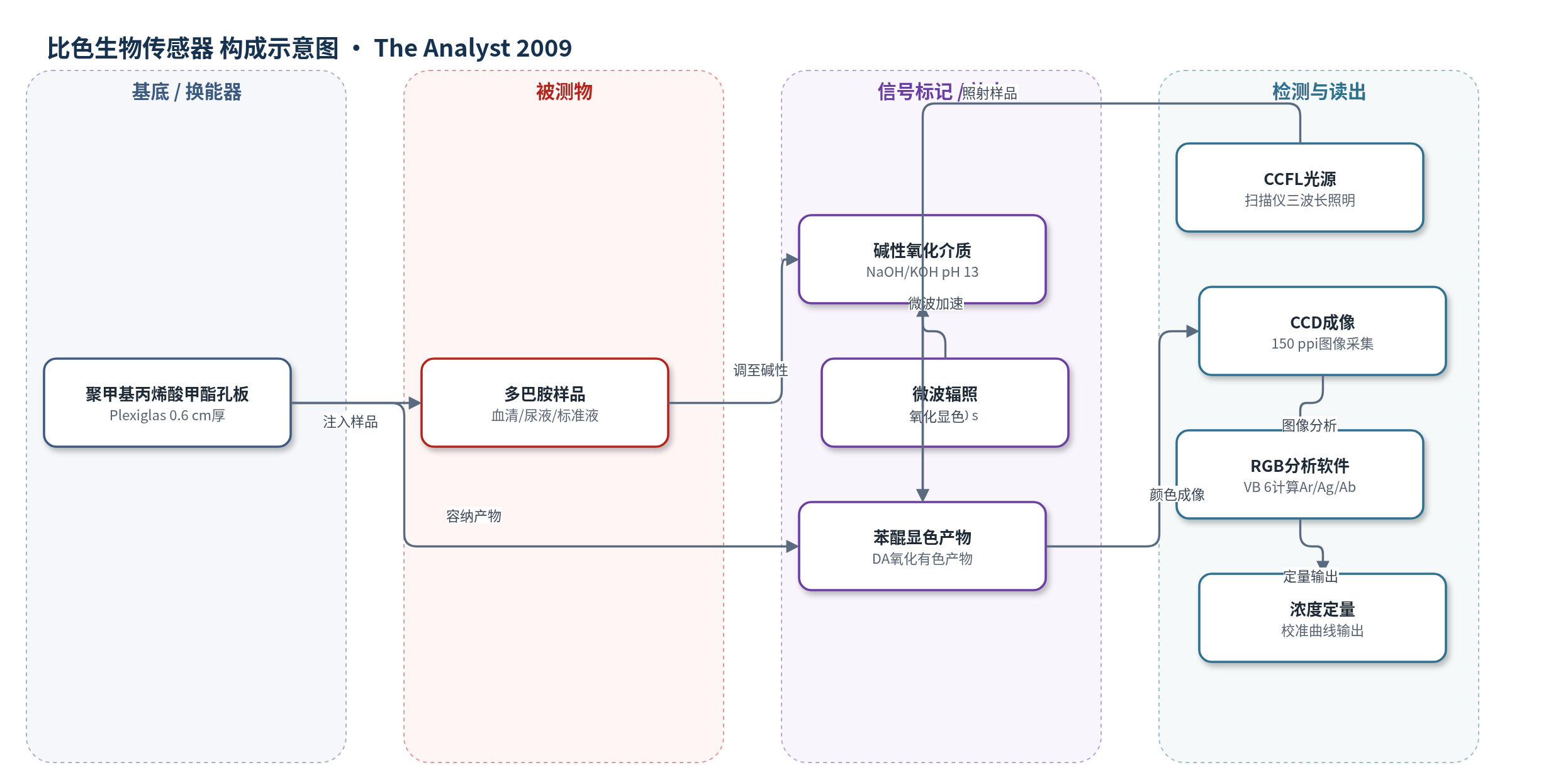
点击图片查看大图 · 依据论文自动绘制
传感器类型
比色生物传感器
检测对象
多巴胺(dopamine, DA);样品基质:牛血清、人尿液、标准溶液
检测原理
多巴胺在强碱性介质(pH 13,NaOH/KOH)中被溶解氧氧化,生成苯醌类有色产物;氧化产物颜色强度随多巴胺浓度增加而增强。样品注入 Plexiglas 孔板并调碱后,可经 150 W 微波辐照 10 s 加速反应。扫描仪 CCFL 光源照射孔板,CCD 采集反射/透射颜色图像,VB 6 软件提取选定区域像素的 RGB 值,并按 Ar=-log(Rs/Rb)、Ag=-log(Gs/Gb)、Ab=-log(Bs/Bb) 计算有效强度。有效强度与浓度建立校准曲线,实现定量。该方法无需额外显色试剂,微波辅助可缩短显色时间并提高灵敏度。
检测灵敏度
LOD: 29 mM (R), 18 mM (G), 12 mM (B)(无微波);16 mM (R), 10 mM (G), 7.5 mM (B)(微波);线性范围: 0.03–1 mM
效应效果
方法对 100 倍尿素、葡萄糖、乳糖无明显干扰;5 倍抗坏血酸对 R、G 值产生约 4.5%–5.0% 相对误差,B 值误差约 -7.0% 至 -7.5%。0.5 mM 多巴胺重现性 RSD 为 2.3%–3.5%(微波)和 2.3%–3.4%(无微波),优于或接近分光光度法(1.8%–2.1%)。牛血清和人尿液加标 5×10^-4 M 后,相对误差约 1.45%–9.20%,无需前处理。与传统 UV-Vis 相比趋势一致,但分光光度法 LOD 和 RSD 更优;微波处理使灵敏度提高近 2 倍,显色时间由 40 min 缩短至 20 min。作者认为其适合低成本商用光学传感。
传感器的构成
- 样品池/反应基底:Plexiglas 激光打孔板(0.6 cm 厚)与 0.1 cm 厚 Plexiglas 封底,形成 21 个圆柱孔,容纳样品溶液。
- 光源:CanoScan 4200F 扫描仪冷阴极荧光灯(CCFL),提供红、绿、蓝三波长照明。
- 检测器:扫描仪电荷耦合器件(CCD),采集孔板颜色图像,分辨率 150 ppi。
- 信号处理:VB 6 软件,将图像像素转换为 RGB 值并计算有效强度 Ar、Ag、Ab。
- 反应介质:NaOH 或 KOH 碱性溶液,将 pH 调至 13,使多巴胺被溶解氧氧化显色。
- 可选加速模块:家用微波炉(Moulinex,150 W,10 s),加速氧化显色并提高灵敏度。
中文摘要
本研究开发了一种简单、快速、低成本的方法,作为可见分光光度法的替代方案。该方法将装有样品溶液的孔板用扫描仪扫描,再用 Visual Basic 6(VB 6)编写的软件将每个孔的颜色解析为红、绿、蓝(RGB)值。孔板由聚甲基丙烯酸甲酯(Plexiglas)板激光打孔制成,并用已知浓度的 Cr(III) 溶液检验各孔尺寸一致性。作者以多巴胺(DA)检测验证方法可行性,检测过程无需其他试剂,并优化了影响体系的参数。与传统 UV-Vis 分光光度法比较,两种方法呈现相似趋势。该方法成功应用于血清和尿液中多巴胺的直接检测,无需样品前处理。结果表明,对溶液进行微波辐照可缩短实验时间、提高灵敏度并改善检出限。
英文摘要
In this study, a new, simple, fast and inexpensive method as an alternative to visible spectrophotometry is developed. In this method the cells containing the sample solution were scanned with a scanner, then the color of each cell was analyzed with software written in visual basic (VB 6) media to red, green and blue values. The cells were built by creating holes in the Plexiglas sheet. The dimensions of identical cells were examined by Cr (III) solution with known concentrations. The validity of this new method was studied by determination of dopamine (DA) without using any other reagent. The parameters which affect the system were optimized. The comparison between the current and traditional UV-Vis spectrophotometry methods was studied and the results revealed similar trends in both methods. The developed method was successfully applied to the determination of dopamine in serum and urine without using any pretreatment. Finally comparing the results obtained in the developed method showed that microwave irradiation of the solution can decrease the experimental time, increase sensitivity and improve the limit of detection.