组成图示
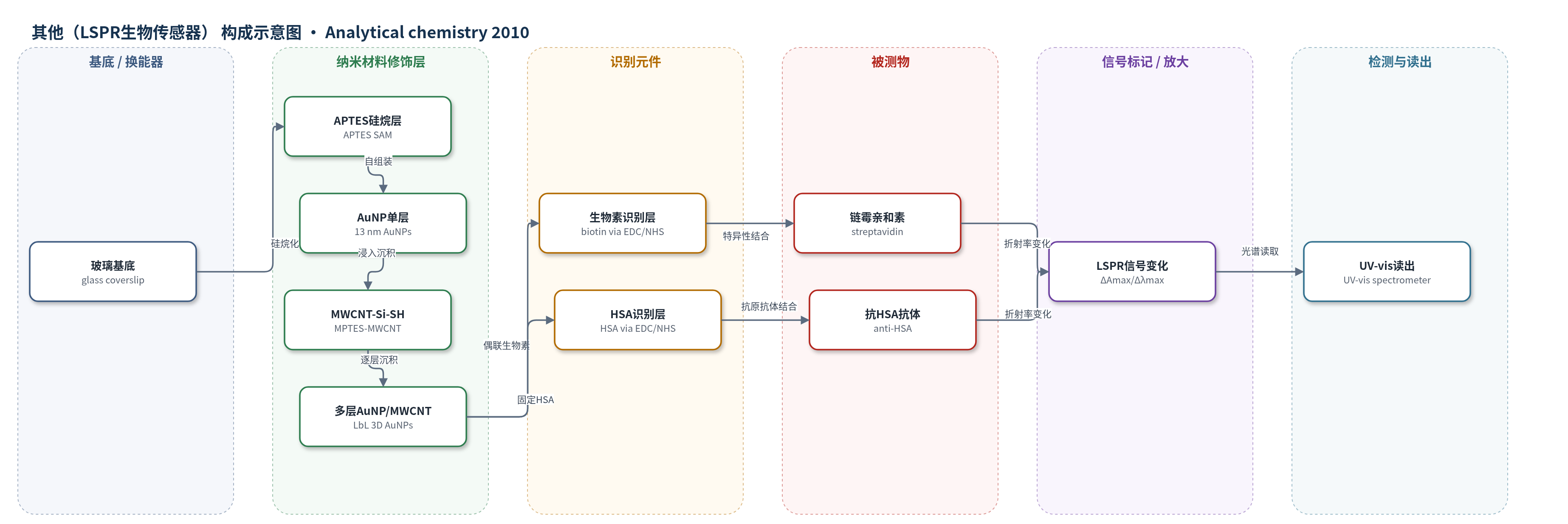
点击图片查看大图 · 依据论文自动绘制
传感器类型
其他(LSPR生物传感器)
检测对象
链霉亲和素(streptavidin,PBS 溶液)、抗人血清白蛋白抗体(anti-HSA,PBS/腹水溶液)
检测原理
玻璃基底经 APTES 硅烷化后自组装 13 nm AuNPs,再逐层沉积 MPTES 功能化 MWCNTs 与 AuNPs,形成三维多孔 AuNP 薄膜。AuNPs 在入射光下产生局域表面等离子共振(LSPR),其消光峰位置和强度对周围介质折射率敏感。当链霉亲和素与表面生物素结合,或抗 HSA 抗体与固定 HSA 结合时,界面附近介电环境/折射率增加,导致 LSPR 峰红移和吸光度增强。三维结构提高 AuNP 密度、结合位点和目标分子可及性,使信号放大。用 UV-vis 记录 ΔAmax(或 Δλmax),随分析物浓度增加而增大。
检测灵敏度
LOD: 0.03 µg/mL (0.5 nM) streptavidin;线性范围: 0.1-25 µg/mL;R^2 = 0.9945;LOD: 0.5 µg/mL (∼3.33 nM) anti-HSA;RI灵敏度: 1.5925 AU/RIU (ΔAmax)、61.29 nm/RIU (Δλmax);2D对照 LOD: 0.6 µg/mL (10 nM) streptavidin、10 µg/mL (∼66.7 nM) anti-HSA;2D RI灵敏度: 0.2853 AU/RIU (ΔAmax)、53.85 nm/RIU (Δλmax)
效应效果
该三维芯片在体折射率响应中比二维 AuNP 单层高 5.6 倍,生物分子检测灵敏度提高约 20 倍。链霉亲和素检测线性范围 0.1–25 µg/mL,R^2=0.9945,LOD 0.03 µg/mL(0.5 nM);抗 HSA 抗体 LOD 0.5 µg/mL(约 3.33 nM),均优于二维芯片的 10 nM 和约 66.7 nM。生物素饱和链霉亲和素对照无吸光度变化,说明信号来自特异性识别。超过三层后重现性下降,因此选择三层作为灵敏度与重现性平衡。未报告实际样品回收率、长期稳定性或 RSD。作者认为该芯片可用于微阵列 LSPR 传感,解决常规 UV-vis 下纳米结构密度低、信号弱的问题。
传感器的构成
- 基底:玻璃盖玻片(glass coverslip),提供透明支撑与 UV-vis 透射路径
- 硅烷化层:APTES(3-aminopropyltriethoxysilane)自组装单层,提供氨基端以固定 AuNPs
- AuNP单层:13 nm AuNPs 自组装单层,形成二维 LSPR 换能表面
- MWCNT功能化层:MWCNT-COOH 与 MPTES 反应生成 MWCNT-Si-SH,引入多个巯基
- 三维复合层:MWCNT-Si-SH 与 AuNPs 逐层沉积形成多层 MWCNT-AuNP,提高 AuNP 密度和微孔可及性
- 识别层(生物素体系):11-AUT SAM 经 EDC/NHS 偶联生物素(biotin),用于捕获链霉亲和素
- 识别层(HSA体系):11-MUA SAM 经 EDC/NHS 固定 HSA,用于结合抗 HSA 抗体
- 信号读出:无标记 LSPR 消光峰强度/波长变化,以 ΔAmax 为主,用 UV-vis 光谱仪读取
中文摘要
通过逐层沉积金纳米颗粒(AuNPs)和多壁碳纳米管(MWCNTs)在玻璃基底上构建三维金纳米结构,用于基于常规紫外-可见光谱仪的高灵敏等离子体生物传感器。羧基化 MWCNTs 与 3-巯基丙基三乙氧基硅烷(MPTES)反应,在 MWCNTs 上引入多个巯基。玻璃芯片上的 AuNP 自组装单层依次浸入 MPTES 功能化 MWCNTs(MWCNT-Si-SH)和 AuNPs,形成 MWCNTs 上的 AuNP 多层结构。这种三维组装 AuNPs 在少数修饰步骤内提供大表面积和多个结合位点,并且多层 MWCNTs 的微孔结构使目标分子具有高可及性。结果表明,这些多层 AuNPs(三维芯片)的体折射率灵敏度比玻璃芯片上 AuNP 单层(二维芯片)高 5.6 倍。三维芯片进一步用于生物分子结合研究,链霉亲和素检出限低至 0.5 nM,抗人血清白蛋白(HSA)抗体检出限为 3.33 nM,均比二维芯片灵敏度高约 20 倍。
英文摘要
Three-dimensional gold nanoarchitecture was fabricated by layer-by-layer (LbL) deposition of gold nanoparticles (AuNPs) and multiwalled carbon nanotubes (MWCNTs) on a glass substrate for a highly sensitive plasmonic biosensor using a conventional UV-vis instrument. Carboxyl-functionalized MWCNTs were reacted with 3-mercaptopropyltriethoxysilane (MPTES) to introduce multiple thiol groups onto MWCNTs. A self-assembled monolayer (SAM) of AuNPs on a glass chip was sequentially dipped into MPTES-functionalized MWCNTs (MWCNT-Si-SH) and AuNPs to form multilayers of AuNPs on MWCNTs. Such three-dimensionally assembled AuNPs provided a large surface area and multiple binding sites within a few steps of modification and microporous structures of multilayered MWCNTs to allow a high accessibility of target molecules. It was shown that the bulk refractive index (RI) sensitivity of these multilayered AuNPs (three-dimensional chip) appeared to be 5.6 times better than that of a monolayer of AuNPs on a glass chip (two-dimensional chip). The three-dimensional chips were further used for a biomolecular binding study, showing a detection limit as low as 0.5 nM for streptavidin and 3.33 nM for anti-human serum albumin (HSA), both of which were approximately 20 times higher than the sensitivity of the two-dimensional chips.