组成图示
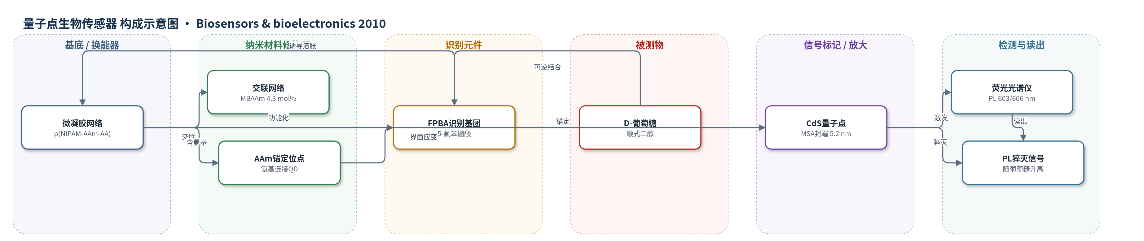
点击图片查看大图 · 依据论文自动绘制
传感器类型
量子点生物传感器
检测对象
葡萄糖(D-glucose/glucose);样品基质为磷酸盐缓冲液(PBS, pH 7.38),并模拟血清干扰物乳酸(l-lactate)和人血清白蛋白(HSA)
检测原理
传感器以 p(NIPAM–AAm–FPBA) 微凝胶为葡萄糖响应基质,FPBA 基团与葡萄糖的顺式二醇形成可逆硼酸酯,使 FPBA 电离度升高并产生 Donnan 电位,驱动微凝胶溶胀。溶胀改变嵌入 CdS 量子点周围的局部折射率和聚合物–QD 界面弹性张力,在界面形成猝灭态,使 QD 荧光强度下降。葡萄糖浓度升高时,微凝胶溶胀程度增大,PL 猝灭增强;在 0–10.8 mM 范围内近似线性,检测限约 0.5 mM,10.8–24.3 mM 在倒数空间呈线性。去除葡萄糖后硼酸酯解离,微凝胶收缩,荧光恢复。该体系无需酶催化,依靠微凝胶体积相变放大荧光变化。
检测灵敏度
LOD: 约0.5 mM;线性范围: 0–10.8 mM;10.8–24.3 mM 倒数空间线性;高交联(6.8%) LOD: 约0.9 mM;低交联(3.2%) LOD: 约0.3 mM
效应效果
在 25 mM 葡萄糖下,原位和呼吸装载杂化微凝胶的 PL 猝灭分别达 65% 和 56%;25 mM 乳酸仅引起约 15% 和 10% 猝灭,2.0 mM 乳酸使葡萄糖响应变化仅 1–3%,表明对乳酸抗干扰良好。44 g/L HSA 在 ≤13.5 mM 葡萄糖下使猝灭增加 <5%,高浓度下因竞争结合使响应不趋于平台。原位微凝胶经 5 次葡萄糖加入/透析循环后 PL 可恢复至原始基线 95% 以上且峰形不变;呼吸装载样品因 QD 泄漏而逐渐下降。交联度 4.3%、6.8%、3.2% 对应猝灭 65%、44%、83%,LOD 约 0.5、0.9、0.3 mM。作者认为其非消耗、可微型化,适合生理条件下连续葡萄糖监测。
传感器的构成
- 微凝胶网络/换能基质:p(NIPAM–AAm–AA) 共聚物微凝胶(NIPAM、AAm、AA,交联剂 MBAAm),提供葡萄糖响应溶胀网络并承载 QD。
- 识别元件:FPBA(2-acrylamidomethyl-5-fluorophenylboronic acid)基团,经 EDC 偶联至 AA 羧基,可逆结合葡萄糖顺式二醇并触发溶胀。
- 信号标记物:CdS 量子点(QDs),原位生长或预合成后嵌入微凝胶,作为光学码产生荧光。
- QD 表面封端/稳定层:MSA(mercaptosuccinic acid)封端预合成 CdS QDs,防止聚集并辅助呼吸装载。
- 界面锚定层:AAm 单元中的 –NH2 基团,锚定 CdS QDs 并限制其泄漏。
- 外部介质:PBS(pH 7.38),维持生理 pH 与离子强度,用于荧光猝灭读出。
中文摘要
本文报道了一类在生理 pH 下具有高灵敏度和高选择性的光学葡萄糖纳米生物传感器。作者将作为光学码的荧光 CdS 量子点(QDs)通过原位生长法和呼吸装载法分别嵌入葡萄糖敏感的聚(N-异丙基丙烯酰胺-丙烯酰胺-2-丙烯酰胺甲基-5-氟苯硼酸)共聚物微凝胶中。该聚合物微凝胶可随周围葡萄糖浓度变化而溶胀或收缩,并调节嵌入 QDs 的荧光,从而将生化信号转换为光学信号;葡萄糖浓度升高时微凝胶逐渐溶胀,导致荧光猝灭。有机–无机杂化微凝胶在生理重要葡萄糖浓度范围内对葡萄糖相对于乳酸和人血清白蛋白表现出高选择性。系统研究了该类杂化微凝胶生物传感器的稳定性、可逆性和灵敏度,并发现其性能可通过调节微凝胶交联度进行调控。基于有机–无机杂化微凝胶的光学葡萄糖纳米生物传感器显示出成为第三代荧光生物传感器的潜力。
英文摘要
A new class of optical glucose nanobiosensors with high sensitivity and selectivity at physiological pH is described. To construct these glucose nanobiosensors, the fluorescent CdS quantum dots (QDs), serving as the optical code, were incorporated into the glucose-sensitive poly(N-isopropylacrylamide-acrylamide-2-acrylamidomethyl-5-fluorophenylboronic acid) copolymer microgels, via both in situ growth method and "breathing in" method, respectively. The polymeric gel can adapt to surrounding glucose concentrations, and regulate the fluorescence of the embedded QDs, converting biochemical signals into optical signals. The gradual swelling of the gel would lead to the quenching of the fluorescence at the elevated glucose concentrations. The hybrid microgels displayed high selectivity to glucose over the potential primary interferents of lactate and human serum albumin in the physiologically important glucose concentration range. The stability, reversibility, and sensitivity of the organic-inorganic hybrid microgel-based biosensors were also systematically studied. These general properties of our nanobiosensors are well tunable under appropriate tailor on the hybrid microgels, in particular, simply through the change in the crosslinking degree of the microgels. The optical glucose nanobiosensors based on the organic-inorganic hybrid microgels have shown the potential for a third generation fluorescent biosensor.