组成图示
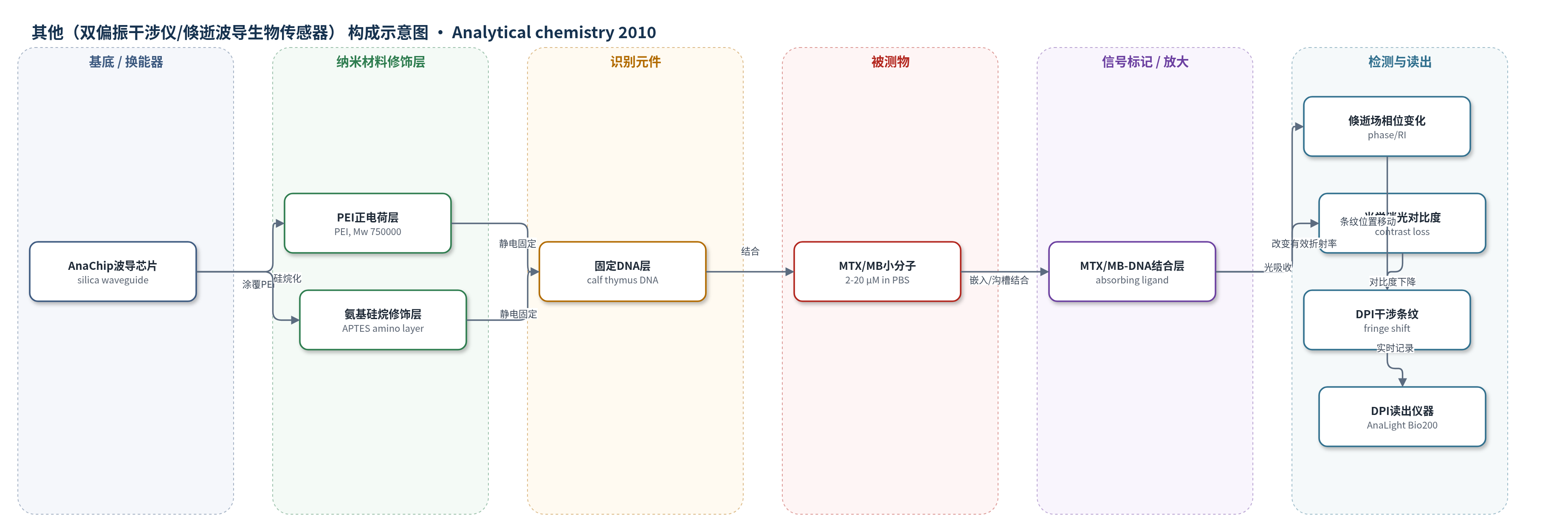
点击图片查看大图 · 依据论文自动绘制
传感器类型
其他(双偏振干涉仪/倏逝波导生物传感器)
检测对象
米托蒽醌(mitoxantrone, MTX)、亚甲蓝(methylene blue, MB)与固定DNA的结合;样品基质:10 mM PBS(pH 7.4,150 mM NaCl)
检测原理
DPI中632.8 nm光在传感与参考波导中传播,倏逝场对界面有效折射率敏感。MTX或MB通过嵌入及沟槽结合与固定DNA结合,结合量随浓度增加而增加。MTX/MB在632.8 nm有强吸收,结合后引起传感波导光强损失,表现为干涉条纹对比度下降,即光学消光,可定量反映小分子结合质量。同时,小分子结合及DNA收缩、致密化、反离子/水分子变化改变有效折射率,使条纹位置移动,即相位变化。相位反映总界面质量与结构变化,光学消光主要反映吸光小分子本身,二者结合可区分结合事件与DNA结构重排。
检测灵敏度
—
效应效果
MTX和MB在DNA表面产生可逆、浓度依赖的光学消光;PEI/氨基排斥表面消光很小,裸芯片仅弱吸附。EtBr嵌入但632.8 nm无吸收,不产生消光,排除散射。光学消光质量给出MTX-DNA Ka=1.8×10^5 M^-1、ka=364 M^-1 s^-1、kd=0.002 s^-1;MB-DNA Ka=4.2×10^4 M^-1、ka=876 M^-1 s^-1、kd=0.021 s^-1。盐浓度影响亲和力:50 mM时MB Ka=9.33×10^4 M^-1,500 mM时1.89×10^4 M^-1。相位显示DNA RI升高、厚度降低,MB厚度变化约为MTX的10倍。与PQCI报道MTX Ka=4.7×10^5 M^-1存在条件差异。方法可实时解析结合与结构变化。
传感器的构成
- 基底/换能器:AnaChip FB80 倏逝波导芯片(silica waveguide),提供传感/参考波导与倏逝场,输出干涉条纹
- 修饰层:PEI(聚乙烯亚胺,Mw 750000)正电荷层,用于在未修饰芯片上静电固定DNA
- 修饰层:氨基硅烷修饰层(3-aminopropyltriethoxysilane/APTES),用于在胺修饰芯片上提供正电荷位点固定DNA
- 识别元件:牛胸腺DNA(calf thymus DNA type I)固定层,作为MTX/MB的结合靶标
- 信号标记物:MTX/MB本身为吸光小分子,结合后在632.8 nm吸收光并改变有效折射率
- 运行缓冲液:10 mM PBS(pH 7.4,150 mM NaCl),维持流动相并调节盐浓度
- 读出系统:DPI仪器AnaLight Bio200,测量相位与对比度/光学消光
中文摘要
本文报道利用双偏振干涉仪(DPI)同时测量光学消光与相位变化,实时探测DNA与小分子相互作用。界面DNA结合米托蒽醌(MTX)和亚甲蓝(MB)后,因光吸收产生可逆、浓度依赖的光学消光,清晰反映小分子与DNA的缔合与解离。基于光学消光推导的质量计算得到MTX-DNA和MB-DNA结合常数分别为1.8×10^5 M^-1和4.2×10^4 M^-1,并显示其受缓冲液盐浓度影响。相位测量反映相互作用总体变化,即小分子结合与DNA结构变化的综合结果;由相位推导的质量可能与光学消光推导的质量显著不同。相位检测到的结构变化表明MTX或MB嵌入后DNA层发生收缩和致密化。光学消光与相位测量相结合可深入理解DNA-小分子相互作用过程。
英文摘要
We demonstrate the use of both optical extinction and phase measurements for probing the interactions between DNA and small molecules by dual polarization interferometry. On binding to DNA at the interface, mitoxantrone (MTX) and methylene blue (MB) induced reversible concentration-dependent optical extinction due to light absorption, which clearly revealed the association and dissociation of small molecules with DNA in real time. The binding constants of MTX-DNA and MB-DNA determined from the masses derived from optical extinction are 1.8 x 10(5) and 4.2 x 10(4) M(-1), respectively, and shown to be buffer salt concentration-dependent. Apart from optical extinction, phase measurements reflected the overall change of the interaction; namely, a combined result of the binding of small molecules and any changes in DNA structure. The masses derived from phase could be very different from those derived from optical extinction. The structural changes detected by phase measurements showed a contraction and densification of DNA upon intercalation by MTX or MB. The combination of optical extinction and phase measurements allows a detailed understanding of the interaction process.