组成图示
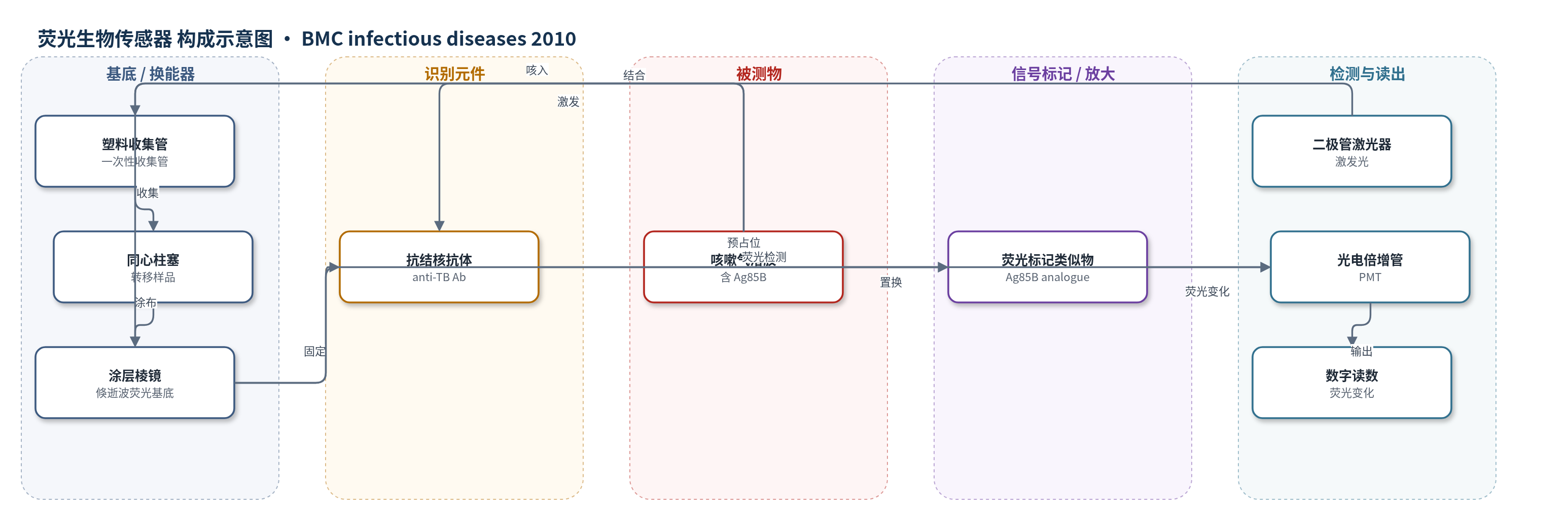
点击图片查看大图 · 依据论文自动绘制
传感器类型
荧光生物传感器
检测对象
结核分枝杆菌抗原85B(Mycobacterium tuberculosis Ag85B),样品基质:咳嗽气溶胶(cough aerosols)
检测原理
该装置采用基于抗原-抗体置换的倏逝波荧光免疫传感原理。一次性收集管底部的棱镜表面固定抗结核抗体,并预先占据荧光标记的 Ag85B 类似物(人工修饰的 T 细胞表位肽亚序列)。患者咳嗽气溶胶中的天然 M. tuberculosis Ag85B 与抗体结合后,将荧光标记类似物从结合位点置换下来,使棱镜表面荧光强度下降。便携式仪器中的二极管激光器激发棱镜表面,产生倏逝波荧光;光电倍增管检测荧光变化,数字模块在约 2 分钟内输出读数。被测抗原越多,荧光信号下降越明显;2 分钟内信号下降超过 20 个单位判为阳性,上升超过 40 个单位判为无效。该法未使用酶催化或核酸扩增放大,主要依靠表面免疫置换和光学荧光读出实现半定量检测。
检测灵敏度
LOD: 未确定,预计为 50–75 colony forming units;判读阈值: 2 min 内信号下降 >20 units 为阳性,上升 >40 units 为无效。
效应效果
现场试点中 60 名呼吸道症状成人均完成检测,过程耐受良好,每人总时间少于 10 分钟。29 例(48.3%)呼吸抗原检测阳性。31 例临床诊断结核患者中,23 例(74%;95% CI 55–87)呼吸抗原阳性,20 例(64%;95% CI 45–80)痰涂片抗酸杆菌阳性;呼吸抗原与涂片镜检联合可检出 29/31(94%;95% CI 77–99)结核患者。6 例非结核患者出现表观假阳性,估计特异性约 79%(95% CI 60–91);另有 6 例涂片阳性结核患者呼吸检测阴性。作者认为该装置不能替代痰涂片镜检,但便携、快速、操作简便,可能用于早期识别传染性结核,尤其适用于无法咳痰人群;一次性装置大规模生产预计成本低于 5 美元。
传感器的构成
- 样品收集层:一次性塑料收集管(plastic collection tube,3.5×10 cm),用于收集咳嗽气溶胶和颗粒。
- 样品转移层:内部同心柱塞(concentric plunger),按压并旋转以将管壁样品转移并涂布至棱镜表面。
- 换能基底:涂层棱镜(coated prism),位于收集管底部,作为检测反应表面并支持倏逝波荧光检测。
- 识别元件:抗结核抗体(anti-tuberculosis antibodies),固定于棱镜表面,用于捕获结核分枝杆菌 Ag85B 抗原。
- 信号标记物:荧光标记类似物(fluorescent-labeled analogue),由人工修饰的 M. tuberculosis Ag85B T 细胞表位肽亚序列组成,预先占据抗体结合位点。
- 光学激发源:二极管激光器(diode laser),提供激发光以产生倏逝波荧光信号。
- 信号读出器:光电倍增管(photo-multiplier tube, PMT)与数字显示模块,检测荧光变化并输出结果。
中文摘要
结核病通过感染者呼吸道气溶胶传播,是高传染性呼吸道疾病。本文报道一种利用免疫传感器与生物光学技术检测咳嗽样本中结核分枝杆菌抗原(Ag85B)的便携式装置,并在高结核发病率地区开展现场试点。TB Breathalyzer 在埃塞俄比亚阿达玛医院门诊用于呼吸道症状成人。患者经 0.9% 生理盐水雾化后向一次性收集管咳嗽,使咳嗽气溶胶沉积;随后将收集管插入便携仪器,判断样本中是否存在抗原。研究记录人口学与临床资料,所有患者均接受胸部 X 线检查和痰液 Ziehl-Neelsen 抗酸染色镜检;缺乏培养时,治疗决策依据涂片镜检、胸片和临床评估。60 名受试者均完成检测,耐受良好,每人少于 10 分钟。29 例(48%)呼吸抗原阳性;31 例临床诊断结核患者中,23 例(74%;95% CI 55–87)呼吸抗原阳性,20 例(64%;95% CI 45–80)痰涂片抗酸杆菌阳性。6 例出现与结核诊断不一致的表观假阳性。作者认为该装置值得作为研究结核分枝杆菌呼出情况的工具进一步验证,其便携、简便、快速的特点提示可能有助于早期识别传染性病例,并建议开展与微生物学及临床指标比较的诊断敏感性和特异性研究。
英文摘要
BACKGROUND: Tuberculosis is a highly infectious disease that is spread from person to person by infected aerosols emitted by patients with respiratory forms of the disease. We describe a novel device that utilizes immunosensor and bio-optical technology to detect M. tuberculosis antigen (Ag85B) in cough and demonstrate its use under field conditions during a pilot study in an area of high TB incidence.
METHODS: The TB Breathalyzer device (Rapid Biosensor Systems Ltd) was field tested in the outpatient clinic of Adama Hospital, Ethiopia. Adults seeking diagnosis for respiratory complaints were tested. Following nebulization with 0.9% saline patients were asked to cough into a disposable collection device where cough aerosols were deposited. Devices were then inserted into a portable instrument to assess whether antigen was present in the sample. Demographic and clinical data were recorded and all patients were subjected to chest radiogram and examination of sputum by Ziehl-Nielsen microscopy. In the absence of culture treatment decisions were based on smear microscopy, chest x-ray and clinical assessment. Breathalyzer testing was undertaken by a separate physician to triage and diagnostic assessment.
RESULTS: Sixty individuals were each subjected to a breathalyzer test. The procedure was well tolerated and for each patient the testing was completed in less than 10 min. Positive breath test results were recorded for 29 (48%) patients. Of 31 patients with a diagnosis of tuberculosis 23 (74%; 95% CI 55-87) were found positive for antigen in their breath and 20 (64%; 95% CI 45-80) were smear positive for acid fast bacilli in their sputum. Six patients provided apparent false positive breathalyzer results that did not correlate with a diagnosis of tuberculosis.
CONCLUSIONS: We propose that the breathalyzer device described warrants further investigation as a tool for studying exhalation of M. tuberculosis. The portability, simplicity of use and speed of the test device suggest it may also find use as a tool to aid early identification of infectious cases. We recommend studies be undertaken to determine the diagnostic sensitivity and specificity of the device when compared to microbiological and clinical indicators of tuberculosis disease.