组成图示
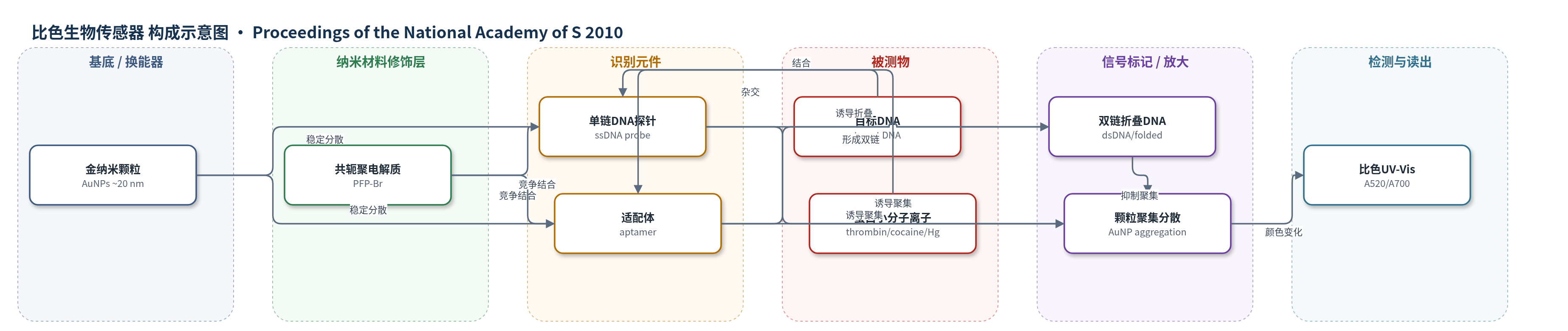
点击图片查看大图 · 依据论文自动绘制
传感器类型
比色生物传感器
检测对象
目标DNA(target DNA)、凝血酶(thrombin)、可卡因(cocaine)、汞离子(Hg(II));样品基质:112.5 mM NaCl缓冲盐溶液及加标血清(blood serum)
检测原理
未修饰金纳米颗粒(AuNPs)在低盐条件下可被单链DNA(ssDNA)或单链适配体稳定,呈520 nm表面等离子共振红色。加入阳离子共轭聚电解质PFP-Br后,其优先结合ssDNA,使ssDNA从AuNPs表面脱离,颗粒聚集,吸收红移,溶液变蓝。若目标DNA与探针杂交形成双链DNA(dsDNA),或凝血酶、可卡因、Hg(II)等目标诱导适配体折叠/缔合为紧凑结构,这些结构结合PFP-Br较弱,仍能稳定AuNPs,溶液保持红色。目标浓度越高,dsDNA/折叠结构越多,A520升高、A700降低,A520/A700增大,颜色越红;无目标则变蓝。该过程无需酶放大或标记,通过肉眼或UV-Vis读出。
检测灵敏度
LOD: 1 pM (naked eyes);LOD: 1.25 pM;<125 attomoles of target in 10 μL solution;thrombin LOD: 10 nM;cocaine LOD: 10 μM;Hg(II) LOD: 50 μM;线性范围: 1.25 pM–125 nM(UV-Vis A520/A700)
效应效果
该比色法在5–10 min内完成,DNA肉眼检测限约1 pM,最低可区分1.25 pM或10 μL中<125 attomoles目标;凝血酶、可卡因、Hg(II)检测限分别为10 nM、10 μM、50 μM。特异性方面,3、5、7碱基错配不能稳定AuNPs而呈蓝色;可卡因传感器对ATP无响应;Exonuclease III预处理可去除双链DNA污染。方法可直接在加标血清中工作。与文献方法相比,本文1 pM优于4.3 nM比色法、100 pM酶放大比色法,接近10 pM电化学法和2.14 pM荧光法,且无需复杂仪器,适合快速现场检测;局限是不适用于有色样品。
传感器的构成
- 换能纳米材料:未修饰金纳米颗粒(unmodified AuNPs,约20 nm),提供520 nm表面等离子共振吸收,聚集后颜色由红变蓝。
- 识别元件:单链DNA探针(ssDNA probe)或适配体(aptamer,如凝血酶适配体、可卡因适配体片段、Hg(II)响应序列),负责与目标特异性结合并改变构象。
- 信号竞争剂:阳离子水溶性共轭聚电解质PFP-Br,优先结合ssDNA,使其脱离AuNPs表面并诱导聚集。
- 稳定/分散介质:112.5 mM NaCl盐溶液,提供低离子强度环境,调节DNA-AuNP静电稳定与聚电解质结合。
- 预处理酶(可选):Exonuclease III,降解样品中双链DNA污染,避免假阳性稳定AuNPs。
- 被测物:目标DNA(target DNA)、凝血酶(thrombin)、可卡因(cocaine)、Hg(II),与探针/适配体结合形成双链或折叠结构。
- 信号读出:肉眼比色或UV-Vis光谱(A520/A700),通过AuNPs分散/聚集状态判断目标存在。
中文摘要
本文报道了一种基于未修饰金纳米颗粒(AuNPs)、单链DNA探针和带正电水溶性共轭聚电解质(PFP-Br)的新型比色传感策略,用于检测DNA序列、蛋白质、小分子和无机离子。该策略利用共轭聚电解质优先结合单链DNA并使其从金纳米颗粒表面脱离,从而诱导AuNPs聚集并发生红-蓝颜色变化;而双链DNA或折叠/缔合DNA与聚电解质结合较弱,可继续稳定AuNPs,使溶液保持红色。基于此,目标DNA与探针杂交形成双链后可抑制聚集,实现肉眼可见的比色检测,检测限达皮摩尔级,并可在血清等复杂基质中工作。进一步利用适配体在目标结合后由单链转变为折叠或双链结构,将该方法推广至凝血酶、可卡因和Hg(II)等分析物检测。该方法操作简便、快速,仅需数分钟即可完成,无需复杂仪器。
英文摘要
We have demonstrated a novel sensing strategy employing single-stranded probe DNA, unmodified gold nanoparticles, and a positively charged, water-soluble conjugated polyelectrolyte to detect a broad range of targets including nucleic acid (DNA) sequences, proteins, small molecules, and inorganic ions. This nearly "universal" biosensor approach is based on the observation that, while the conjugated polyelectrolyte specifically inhibits the ability of single-stranded DNA to prevent the aggregation of gold-nanoparticles, no such inhibition is observed with double-stranded or otherwise "folded" DNA structures. Colorimetric assays employing this mechanism for the detection of hybridization are sensitive and convenient--picomolar concentrations of target DNA are readily detected with the naked eye, and the sensor works even when challenged with complex sample matrices such as blood serum. Likewise, by employing the binding-induced folding or association of aptamers we have generalized the approach to the specific and convenient detection of proteins, small molecules, and inorganic ions. Finally, this new biosensor approach is quite straightforward and can be completed in minutes without significant equipment or training overhead.