组成图示
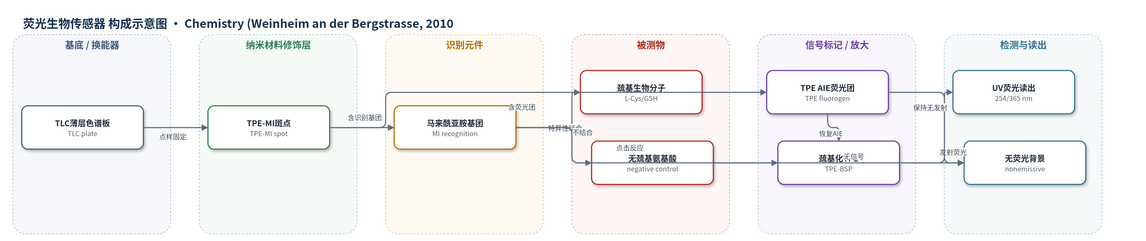
点击图片查看大图 · 依据论文自动绘制
传感器类型
荧光生物传感器
检测对象
L-半胱氨酸(L-cysteine)、谷胱甘肽(GSH)等含自由巯基生物分子;样品基质为 DMSO 溶液及活 HeLa 细胞培养体系
检测原理
TPE-MI 中马来酰亚胺(MI)单元的羰基与烯键形成 n-π 共轭,导致 TPE 的荧光在溶液和固态均被淬灭。当含自由巯基的分析物(如 L-半胱氨酸、GSH)接触 TLC 板上的 TPE-MI 斑点时,巯基与 MI 发生巯基-烯点击加成,破坏该共轭并生成巯基化 TPE(TPE-BSP)。由于 TPE 具有聚集诱导发光(AIE)特性,反应产物在固态/聚合态下恢复强蓝色荧光。荧光强度随巯基浓度增加而增强,低浓度约 1 ppb 时仍可肉眼观察;不含自由巯基的氨基酸或蛋白不反应,斑点保持暗态。在活细胞中,TPE-MI 纳米聚集体进入细胞并与胞内巯基(主要为 GSH)反应,从而以荧光信号映射巯基分布。
检测灵敏度
LOD: 1 ng/mL(约 1 ppb,L-cysteine,TLC 板肉眼检测);GSH: 约 0.3 ppm
效应效果
该探针具有高度选择性,仅响应自由巯基:L-半胱氨酸(L-cysteine)在 1–1000 ng/mL 范围内均产生可见荧光,1 ng/mL(约 1 ppb)仍可肉眼辨识;而天冬氨酸、亮氨酸、苯丙氨酸、精氨酸、天冬酰胺、蛋氨酸和谷氨酸等无巯基氨基酸均不发光。谷胱甘肽(GSH)约 0.3 ppm 即可开启发射,无巯基蛋白 GLVPA 无响应。TPE-MI 纳米聚集体孵育 HeLa 细胞 5 min 后,细胞保持健康,显示良好生物相容性;荧光主要位于胞质,核区较弱。方法无需昂贵仪器,操作简便、快速,适合固态巯基检测与细胞内 GSH 分布成像。
传感器的构成
- 基底/固态载体:薄层色谱板(TLC plate),提供固态反应与成像平台
- 荧光探针层:四苯乙烯-马来酰亚胺(TPE-MI)斑点,TPE 为 AIE 荧光团,MI 为识别基团
- 识别元件:马来酰亚胺(MI)基团,通过巯基-烯点击反应特异性结合自由巯基
- 信号换能元件:TPE 聚合态 AIE 荧光体,巯基加成后恢复强蓝色荧光
- 读出层:254/365 nm UV 激发下肉眼或荧光成像观察斑点发光
中文摘要
本文报道了一种便捷、特异且灵敏的固态荧光生物探针。作者将四苯乙烯(TPE)通过马来酰亚胺(MI)基团功能化,得到 TPE-MI 加合物;该加合物在溶液和固态下均无荧光发射。当含自由巯基的分析物存在时,巯基与 MI 侧基发生巯基-烯点击加成,破坏 MI 单元的 n-π 共轭,使 TPE 恢复聚集诱导发光(AIE)性质,从而由暗态转变为强蓝色荧光。基于该机制,TPE-MI 斑点在薄层色谱(TLC)板上暴露于 L-半胱氨酸等含巯基氨基酸时发光,而暴露于其他无自由巯基氨基酸时保持无发射。该过程快速、对比度高,约 1 ppb 的 L-半胱氨酸即可被肉眼识别。类似地,含自由巯基的蛋白质如谷胱甘肽(GSH)也能开启 TPE-MI 的发射。以 TPE-MI 纳米聚集体作为可视化剂对活细胞染色后,可获得清晰荧光图像,为细胞系统中巯基物种分布的简便荧光标记提供了工具。
英文摘要
A handy, specific, sensitive bioprobe has been developed. Tetraphenylethene (TPE) was functionalized by a maleimide (MI) group, giving a TPE-MI adduct that was nonemissive in both solution and the solid state. It was readily transformed into a fluorogen showing an aggregation-induced emission (AIE) property by the click addition of thiol to its MI pendant. The click reaction and the AIE effect enabled TPE-MI to function as a thiol-specific bioprobe in the solid state. Thus, the spot of TPE-MI on a TLC plate became emissive when it had been exposed to L-cysteine, an amino acid containing a thiol group, but remained nonemissive when exposed to other amino acids that lack free thiol units. The thiol-activated emission was rapid and strong, readily detected by the naked eye at an analyte concentration as low as approximately 1 ppb, thanks to the "lighting up" nature of the bioprobing process. Similarly, the emission of TPE-MI was turned on only by the proteins containing free thiol units, such as glutathione. Clear fluorescence images were taken when living cells were stained by using TPE-MI as a visualization agent, affording a facile fluorescent maker for mapping the distribution of thiol species in cellular systems.