组成图示
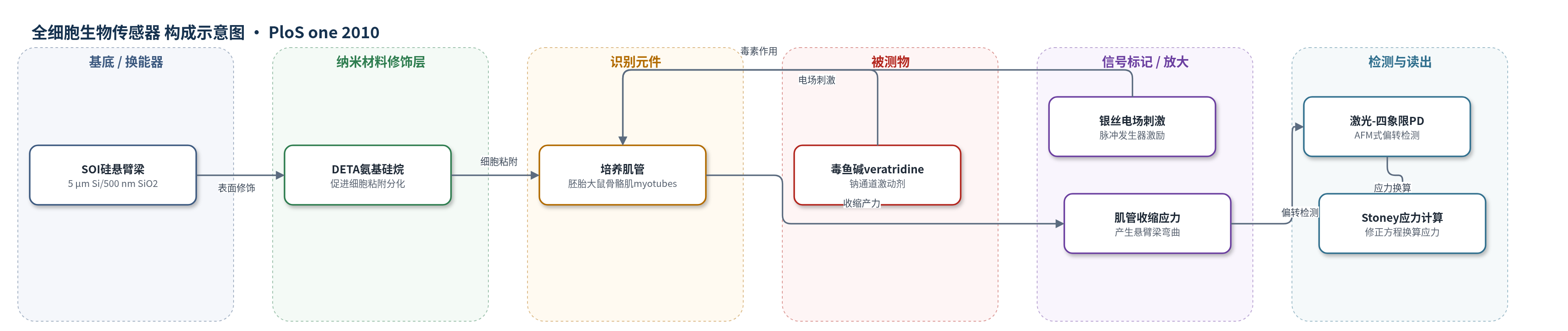
点击图片查看大图 · 依据论文自动绘制
传感器类型
全细胞生物传感器
检测对象
毒鱼碱(veratridine)、肌管收缩应力(contractile stress);样品基质:培养于硅悬臂梁上的胚胎大鼠骨骼肌肌管及Neurobasal/B-27或NbActiv4培养基。
检测原理
该装置以培养肌管作为生物识别与效应元件,DETA修饰的硅悬臂梁提供粘附界面。电场刺激或毒鱼碱作用于肌管膜上的电压门控钠通道,引起钠离子内流、肌浆网钙释放和肌管收缩。收缩在悬臂梁表面形成薄膜应力,使硅梁发生弯曲;AFM式激光-四象限光电探测器测量末端偏转和角度,进而得到曲率。利用修正Stoney方程、Atkinson近似和Klein校正将偏转换算为应力。毒鱼碱使钠通道持续开放,导致异步强直收缩,峰值应力升高后细胞失能,信号变化反映毒素作用;理论上毒素浓度越高,钠通道开放和收缩响应越强。
检测灵敏度
原文未报告LOD、线性范围、灵敏度斜率或相关系数。
效应效果
装置可实时记录培养肌管收缩,悬臂梁平均厚5.27±0.07 μm,弹簧常数实验值1.27±0.06 N/m,谐振频率14.4±0.3 kHz。Neurobasal/B-27培养肌管峰值应力约0.3–1.1 kPa,TPT 236.8±26.1 ms,KRT 233.6±23.8 ms,ds/dt 2.4 Pa/ms;NbActiv4培养后应力约1.2–1.3 kPa,TPT 172.1±4.7 ms,KRT 175.6±3.6 ms,ds/dt 11.8 Pa/ms,提示更成熟表型。加入5 mM veratridine后出现异步强直收缩,峰值应力显著升高随后回落基线。肌管覆盖率>95%,培养可维持4周乃至90天。作者认为可用于高吞吐肌肉生理、毒理学和药物筛选。
传感器的构成
- 基底/换能器:SOI硅悬臂梁(5 μm Si/500 nm SiO2,DRIE刻蚀,平均厚5.27±0.07 μm),承载肌管并将收缩应力转换为弯曲。
- 表面修饰层:DETA氨基硅烷((3-Trimethoxysilyl propyl) diethylenetriamine),涂覆于硅表面,促进肌细胞粘附、融合与分化。
- 识别/效应元件:胚胎大鼠骨骼肌肌管(myotubes),培养10–13天形成,沿悬臂梁长轴取向,收缩时产生薄膜应力。
- 激励元件:银丝电极(0.015 inch,间距15 mm)与脉冲发生器,用于电场刺激肌管同步收缩。
- 检测读出:AFM式红色激光-四象限光电探测器(PD)系统,测量悬臂梁末端偏转和角度变化。
- 信号处理:修正Stoney方程/Atkinson近似/Klein校正,将偏转/曲率换算为肌管应力。
- 样品/响应介质:Neurobasal/B-27或NbActiv4培养基,veratridine作为外源毒素响应物。
中文摘要
本文报道了一种基于硅悬臂梁和原子力显微镜式光学检测系统的生物微机电系统(Bio-MEMS),用于研究胚胎大鼠骨骼肌来源肌管的生理与动力学。该系统可实时观察肌管行为,并通过电场刺激选择性诱发收缩。利用修正的Stoney方程估算肌管在悬臂梁上产生的应力,所得应力值与已发表的培养肌管结果高度一致,但低于成年骨骼肌水平。与文献相比,培养肌管虽具有可比的峰值应力,但达到峰值张力的时间(TPT)和半弛豫时间(KRT)更长;使用增强培养基NbActiv4培养后,这些参数明显改善,提示肌管呈胚胎表型。此外,系统对钠通道激动剂毒鱼碱(veratridine)表现出响应,加入毒素后肌管出现强直收缩并随后失去收缩能力。作者认为该装置可为高吞吐肌肉生理研究、生物传感器和药物发现提供基础。
英文摘要
BACKGROUND: To date, biological components have been incorporated into MEMS devices to create cell-based sensors and assays, motors and actuators, and pumps. Bio-MEMS technologies present a unique opportunity to study fundamental biological processes at a level unrealized with previous methods. The capability to miniaturize analytical systems enables researchers to perform multiple experiments in parallel and with a high degree of control over experimental variables for high-content screening applications.
METHODOLOGY/PRINCIPAL FINDINGS: We have demonstrated a biological microelectromechanical system (BioMEMS) based on silicon cantilevers and an AFM detection system for studying the physiology and kinetics of myotubes derived from embryonic rat skeletal muscle. It was shown that it is possible to interrogate and observe muscle behavior in real time, as well as selectively stimulate the contraction of myotubes with the device. Stress generation of the tissue was estimated using a modification of Stoney's equation. Calculated stress values were in excellent agreement with previously published results for cultured myotubes, but not adult skeletal muscle. Other parameters such as time to peak tension (TPT), the time to half relaxation ((1/2)RT) were compared to the literature. It was observed that the myotubes grown on the BioMEMS device, while generating stress magnitudes comparable to those previously published, exhibited slower TPT and (1/2)RT values. However, growth in an enhanced media increased these values. From these data it was concluded that the myotubes cultured on the cantilevers were of an embryonic phenotype. The system was also shown to be responsive to the application of a toxin, veratridine.
CONCLUSIONS/SIGNIFICANCE: The device demonstrated here will provide a useful foundation for studying various aspects of muscle physiology and behavior in a controlled high-throughput manner as well as be useful for biosensor and drug discovery applications.