组成图示
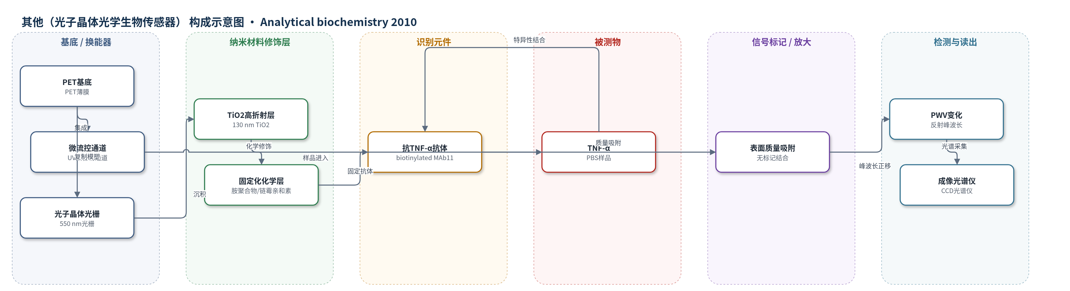
点击图片查看大图 · 依据论文自动绘制
传感器类型
其他(光子晶体光学生物传感器)
检测对象
肿瘤坏死因子-α(TNF-α);样品基质:PBS(磷酸盐缓冲液)
检测原理
TNF-α样品与固定在光子晶体表面的抗TNF-α抗体发生特异性结合,使传感表面质量密度增加。光子晶体由低折射率聚合物光栅和TiO2高折射层构成,表面质量变化会改变倏逝场耦合的有效折射率,使反射共振峰波长值(PWV)向长波方向移动。PWV移动量与结合质量近似相关,原文采用9 pg mm-2对应1 nm PWV的换算关系。微流控格式通过流动增强分析物向表面的质量传输,点状固定化减少非传感表面对分析物的捕获。反射光谱读出PWV变化,浓度越高平衡结合量越大,PWV正移越大;达到平衡的时间受扩散、流速、配体密度和点尺寸影响。
检测灵敏度
LOD: 16.7 ng ml-1 (channel-whole/channel-spot); 30.9 ng ml-1 (well-whole); 48.4 ng ml-1 (well-spot); 质量密度灵敏度: 9 pg mm-2 per 1 nm PWV shift
效应效果
实验与有限元模拟趋势一致,未使用人工缩放参数。50 ng/mL TNF-α下,channel-whole/channel-spot平衡PWV为0.0195 nm,达到平衡时间分别为19.3和10.60 min,LOD为16.7 ng/mL;well-whole平衡PWV为0.0105 nm、时间1965 min、LOD 30.9 ng/mL;well-spot平衡PWV为0.0186 nm、时间498 min、LOD 48.4 ng/mL。PWV标准差:channel-whole/channel-spot为0.00220 nm,well-whole为0.00203 nm,well-spot为0.00601 nm。测得TNF-α/抗TNF-α Kd=0.121±0.0452 μM,与QCM报道0.231±0.542 μM相当。封闭后非特异结合仍可能存在。作者认为微流控格式检测最快、平衡信号最大、LOD最低并可测动力学;well-spot配体消耗最低,适合昂贵抗体;well-whole协议简单、分析物消耗低。
传感器的构成
- 基底/换能器:PET(polyethylene terephthalate)薄膜或PC载玻片,承载光子晶体结构并提供机械支撑。
- 光子晶体换能层:UV固化聚合物(Gelest)一维光栅,周期550 nm、深度170 nm,产生窄带反射峰,表面质量变化引起PWV移动。
- 高折射率敏感层:TiO2(titanium dioxide)130 nm电子束蒸发层,增强倏逝场耦合与表面光学响应。
- 微流控通道层:UV固化聚合物复制模塑通道,深度30 μm,用于输送TNF-α样品与缓冲液。
- 化学固定化层:胺聚合物(amine polymer)、戊二醛(glutaraldehyde)和链霉亲和素(streptavidin)构建抗体固定界面;well-spot使用环氧硅烷(3-glycidoxypropyldimethylethoxysilane)形成疏水点状固定表面。
- 识别元件:生物素化抗人TNF-α单克隆抗体(biotinylated anti-human TNF-α MAb11)或anti-TNF-α抗体,全表面或点状固定,特异性捕获TNF-α。
- 封闭剂:BioFX milk blocker封闭未结合抗体区域,降低非特异结合。
- 信号读出层:白光/近红外LED、成像光谱仪或光纤光谱仪、CCD,测量反射峰波长值(PWV)变化。
中文摘要
本研究结合实验检测与流体动力学有限元模拟,直接比较了四种无标记生物传感检测配置的性能极限:将光子晶体生物传感器集成于微孔板孔底或微流控通道中,并分别比较捕获配体全表面涂覆与局部点状固定两种情形。以肿瘤坏死因子-α(TNF-α)为模型分析物,采用固定化抗TNF-α捕获抗体进行蛋白-抗体结合检测。结果表明,微流控检测格式可显著缩短达到稳定平衡所需时间;对于微孔板检测,点状固定化有利于减少达到平衡响应所需时间。所建立模型和结论适用于任意无标记生物传感器技术以及任意配体-分析物体系,可调变量包括传感器质量密度灵敏度、分析物-配体吸附/解离速率常数、固定化配体密度、流道几何、流速和点尺寸。
英文摘要
Using both experimental assays and fluid-dynamic finite element simulation models, we directly compared the achievable performance limits of four distinct assay configurations for label-free detection of an analyte from a test sample on a biosensor surface. The assay configurations studied in this work included a biosensor incorporated into the bottom surface of a microplate well and a microfluidic channel. For each configuration, we compared assay performance for the scenario in which the entire bottom surface of the fluid-handling vessel is coated with capture ligands with assay performance for the scenario in which the capture ligands are applied in the form of localized spots. As a model system, we used detection of the protein biomarker tumor necrosis factor-alpha (TNF-alpha) using immobilized TNF-alpha capture antibody. Results show that the microfluidic assay format dramatically reduces the time required to establish a stable equilibrium. Spot-based assays are advantageous for microplate-based detection for reducing the time required for equilibrium sensor response. The results derived are generally applicable to any label-free biosensor technology and any ligand-analyte system with adjustable variables that include sensor mass density sensitivity, analyte-ligand adsorption/desorption rate constants, immobilized ligand density, flow channel geometry, flow rate, and spot size.