组成图示
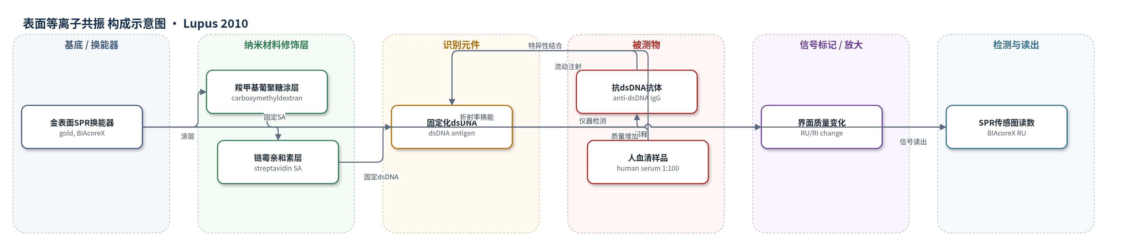
点击图片查看大图 · 依据论文自动绘制
传感器类型
表面等离子共振(SPR)生物传感器
检测对象
抗双链DNA抗体(anti-dsDNA);样品基质:人血清(human serum)
检测原理
该SPR生物传感器以金表面为换能基底,经羧甲基葡聚糖涂层和链霉亲和素(SA)固定化dsDNA抗原。人血清稀释后流经芯片,其中抗dsDNA抗体与固定化dsDNA发生特异性结合。结合事件使传感器界面质量浓度增加,引起局部折射率变化;SPR换能器将这种变化转换为共振单位(RU)随时间变化的传感图。作者取结合相最大RU作为测量值,并用抗dsDNA单克隆抗体dsDNA-mAb32校准为IU/ml。信号强度随血清中抗dsDNA抗体浓度及亲和力升高而增大,解离相可反映结合稳定性。该方法无酶促放大,但通过实时监测结合/解离和可再生表面实现重复检测。
检测灵敏度
未报告LOD、线性范围、灵敏度斜率或相关系数。
效应效果
五种方法总体诊断效率均满意。全部SLE与对照中,Bindazyme AUC 0.885、DE 0.88,ELiA AUC 0.886、DE 0.86,SPR AUC 0.820、DE 0.80,Farr AUC 0.770、DE 0.70;Farrzyme DE 0.77。狼疮肾炎亚组AUC为Bindazyme 0.979、ELiA 0.945、SPR 0.928、Farr 0.886、Farrzyme 0.879。SPR用90 µl 1:100血清可产生约100–300 RU,前期25 RU界值特异性98%、敏感性83%;芯片约60循环衰减<8%,校准物305±24 RU。方法间Spearman rs 0.38–0.71(p<0.01)。作者认为SPR可实时监测结合,优化后有助于SLE诊断与监测。
传感器的构成
- 基底/换能器:金表面(gold surface)与BIAcoreX SPR换能器,提供表面等离子共振检测界面
- 修饰层:羧甲基葡聚糖涂层(carboxymethyldextran coating),覆盖金表面并用于固定化介导
- 固定介导层:链霉亲和素(streptavidin, SA)预固定于FF1/FF2芯片,用于固定dsDNA抗原
- 识别元件:双链DNA(dsDNA)抗原,芯片固定,特异性结合血清中抗dsDNA抗体
- 运行缓冲液:0.01 M HEPES pH 7.4、0.15 M NaCl、0.1%牛血清白蛋白(BSA)、0.005%辛基-β-D-葡萄糖苷,维持结合条件并降低非特异吸附
- 校准物:人抗dsDNA单克隆抗体dsDNA-mAb32,用于将RU信号校准为IU/ml
- 再生液:50 mM NaOH/1 M NaCl,用于结合后表面再生
中文摘要
系统性红斑狼疮(SLE)患者常出现多种血清学异常,其中抗双链DNA(anti-dsDNA)抗体阳性是SLE诊断的实验室标准之一,具有重要临床意义。本研究回顾性分析50例临床确诊SLE患者、39例其他自身免疫病患者和20例健康对照,比较四种商业anti-dsDNA免疫测定与一种新建立的表面等离子共振(SPR)生物传感器芯片的诊断准确性。该芯片将dsDNA共价固定于芯片表面。采用受试者工作特征(ROC)曲线分析,并特别关注狼疮肾炎患者。结果显示,四种免疫测定与SPR生物传感器的曲线下面积(AUC)和诊断效率(DE)依次为:Bindazyme AUC 0.89、DE 0.88;ELiA AUC 0.89、DE 0.86;SPR AUC 0.82、DE 0.80;Farrzyme AUC 0.77、DE 0.77;Farr AUC 0.77、DE 0.70。在22例狼疮肾炎SLE患者中,AUC分别为Bindazyme 0.98、ELiA 0.95、SPR 0.93、Farr 0.89、Farrzyme 0.88。尽管方法学不同,所有免疫测定的总体诊断准确性均令人满意,Bindazyme表现最佳。作者以自研SPR生物传感器为参照,其AUC和DE值良好;经优化后,该技术可实时监测抗原抗体相互作用,可能为SLE诊断和临床监测提供新的分析质量。
英文摘要
Patients with systemic lupus erythematosus (SLE) often develop a wide variety of serological manifestations including the presence of antibodies to double-stranded DNA (anti-dsDNA). Positivity for anti-dsDNA constitutes one of the laboratory criteria for the diagnosis of SLE and is therefore clinically relevant. We analyzed the diagnostic accuracies of four commercial anti-dsDNA immunoassays and compared the results with a recently established surface plasmon resonance (SPR) biosensor chip with covalently chip-immobilized dsDNA. The anti-dsDNA measurements were performed retrospectively in 50 patients with clinically proven SLE, 39 patients with other autoimmunopathies and 20 healthy controls. Data were evaluated by Receiver-Operator Characteristic (ROC) analysis, with special regard to SLE patients suffering from lupus nephritis. The ROC analyses for the four immunoassays and the SPR biosensor resulted in the following area-under-the-curve (AUC) and diagnostic efficiency (DE) values in descending order: Bindazyme AUC, 0.89; DE, 0.88; ELiA AUC, 0.89; DE, 0.86; SPR biosensor AUC, 0.82; DE, 0.80; Farrzyme AUC, 0.77; DE, 0.77; Farr AUC, 0.77; DE, 0.70. When considering the 22 nephritis SLE patients the following AUC were observed: Bindazyme 0.98; EliA 0.95; SPR biosensor 0.93; Farr 0.89; Farrzyme 0.88. Although various methodologies for the determination of anti-dsDNA were compared, the overall diagnostic accuracy was found satisfactory in all immunoassays. Best data were found for the Bindazyme assay. We referenced the measurements to our in-house SPR biosensor device which showed good AUC and DE values. When optimized, this technique, allowing to monitor antigen/ antibody interactions in real-time, may add a new analytical quality to the existing methods, potentially beneficial in diagnosis and clinical monitoring of SLE.