组成图示
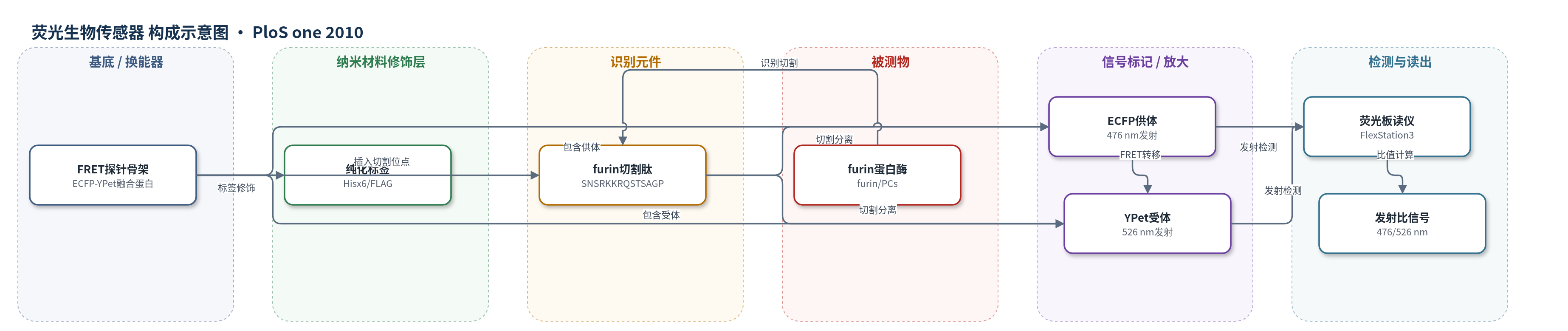
点击图片查看大图 · 依据论文自动绘制
传感器类型
荧光生物传感器
检测对象
furin(furin,前蛋白转化酶活性);样品基质:体外纯化furin、肿瘤细胞表面/细胞培养上清、细胞裂解液(MCF-7、LoVo、U251等)
检测原理
该传感器为切割激活型FRET探针:ECFP作为供体,YPet作为受体,二者通过源自炭疽PA83的furin切割肽SNSRKKRQSTSAGP连接。未切割时,供体与受体距离接近且取向合适,ECFP激发后能量通过FRET转移至YPet,YPet 526 nm发射增强而ECFP 476 nm发射受抑制。当furin识别并切割该多碱性序列后,ECFP与YPet分离,FRET效率下降,ECFP发射增强、YPet发射减弱,lex=437 nm下ECFP/YPet发射比升高。信号变化与furin浓度或活性呈比例关系,因此可定量监测furin活性。选择性主要来自PA83位点对furin/前蛋白转化酶的特异性切割;WNV NS2B-NS3等蛋白酶不切割该探针,非PC蛋白酶即使破坏探针也不特异性切割linker,干扰较低。无酶促沉积或核酸放大,信号放大依赖FRET比值读出。
检测灵敏度
可检测低至 10 fmol furin(原文:levels of furin as low as 10 fmol were sufficient to cause measurable changes)
效应效果
传感器对furin及furin-like前蛋白转化酶敏感,抵抗WNV NS2B-NS3蛋白酶,切割特性与PA83一致;dec-RVKR-cmk可抑制信号,aprotinin不抑制furin。MCF-7:furWT 2 h即可激活,天然表达细胞需8–16 h;MCF-7:furD153N和LoVo不激活。估算5×10^4个MCF-7:furWT细胞含约10 fmol(<0.6 ng,约100,000分子/细胞)furin,U251更低。细胞裂解液中MCF-7:furWT、LoVo:furWT、U251分别约109、43、24 fmol,且可被dec-RVKR-cmk抑制。表面生物素化和抗体摄取显示细胞表面furin极低。与传统荧光肽底物Pyr-RTKR-AMC相比,传感器可在粗细胞裂解液中可靠、连续监测furin活性,适用于肿瘤细胞furin监测和炭疽PA83胞外加工研究。
传感器的构成
- 探针骨架:ECFP-YPet融合蛋白,构成FRET供受体对,是信号换能主体。
- 供体荧光蛋白:ECFP(Enhanced Cyan Fluorescent Protein),作为FRET供体,切割后476 nm发射增强。
- 受体荧光蛋白:YPet(YFP变体),作为FRET受体,切割后526 nm发射降低。
- 识别/切割元件:PA83衍生furin切割肽序列SNSRKKRQSTSAGP,连接ECFP与YPet,被furin/前蛋白转化酶特异性切割。
- 纯化标签:N端Hisx6和FLAG标签,用于Co2+亲和层析纯化,不直接参与FRET信号。
- 信号读出:FlexStation3荧光板读仪,lex=437 nm,测量ECFP/YPet发射比(476 nm/526 nm)。
中文摘要
Furin是一种特异性内蛋白酶,可将前蛋白加工为活性蛋白,在正常细胞及肿瘤、炭疽等疾病中发挥重要作用。Furin被认为在高尔基体与细胞表面之间循环,而炭疽保护性抗原PA83被细胞加工常被视为细胞表面存在大量furin的证据。为监测furin,作者设计了切割激活型FRET生物传感器,将ECFP与YPet通过源自PA83的肽序列SNSRKKRQSTSAGP连接。由于该序列对furin蛋白水解高度敏感且选择性高,传感器可在体外反应和细胞实验中记录飞摩尔水平的furin。利用因尺寸不能进入细胞的传感器及其他方法,作者发现完整细胞及过表达furin的细胞中,细胞表面furin水平极低甚至几乎不存在,这与既有观点形成鲜明对比。细胞实验表明,PA83实际在胞外环境中被furin加工,随后PA63结合炭疽毒素细胞表面受体。此外,该传感器可连续监测癌细胞裂解液中furin活性。结果提示不存在具有生理意义的细胞表面furin,炭疽机制需重新研究;该传感器也为非侵入监测癌细胞furin活性奠定基础,并可作为其他蛋白酶激活型生物传感器的原型。
英文摘要
Furin, a specialized endoproteinase, transforms proproteins into biologically active proteins. Furin function is important for normal cells and also in multiple pathologies including malignancy and anthrax. Furin is believed to cycle between the Golgi compartment and the cell surface. Processing of anthrax protective antigen-83 (PA83) by the cells is considered thus far as evidence for the presence of substantial levels of cell-surface furin. To monitor furin, we designed a cleavage-activated FRET biosensor in which the Enhanced Cyan and Yellow Fluorescent Proteins were linked by the peptide sequence SNSRKKR / STSAGP derived from anthrax PA83. Both because of the sensitivity and selectivity of the anthrax sequence to furin proteolysis and the FRET-based detection, the biosensor recorded the femtomolar levels of furin in the in vitro reactions and cell-based assays. Using the biosensor that was cell-impermeable because of its size and also by other relevant methods, we determined that exceedingly low levels, if any, of cell-surface furin are present in the intact cells and in the cells with the enforced furin overexpression. This observation was in a sharp contrast with the existing concepts about the furin presentation on cell surfaces and anthrax disease mechanism. We next demonstrated using cell-based tests that PA83, in fact, was processed by furin in the extracellular milieu and that only then the resulting PA63 bound the anthrax toxin cell-surface receptors. We also determined that the biosensor, but not the conventional peptide substrates, allowed continuous monitoring of furin activity in cancer cell extracts. Our results suggest that there are no physiologically-relevant levels of cell-surface furin and, accordingly, that the mechanisms of anthrax should be re-investigated. In addition, the availability of the biosensor is a foundation for non-invasive monitoring of furin activity in cancer cells. Conceptually, the biosensor we developed may serve as a prototype for other proteinase-activated biosensors.