组成图示
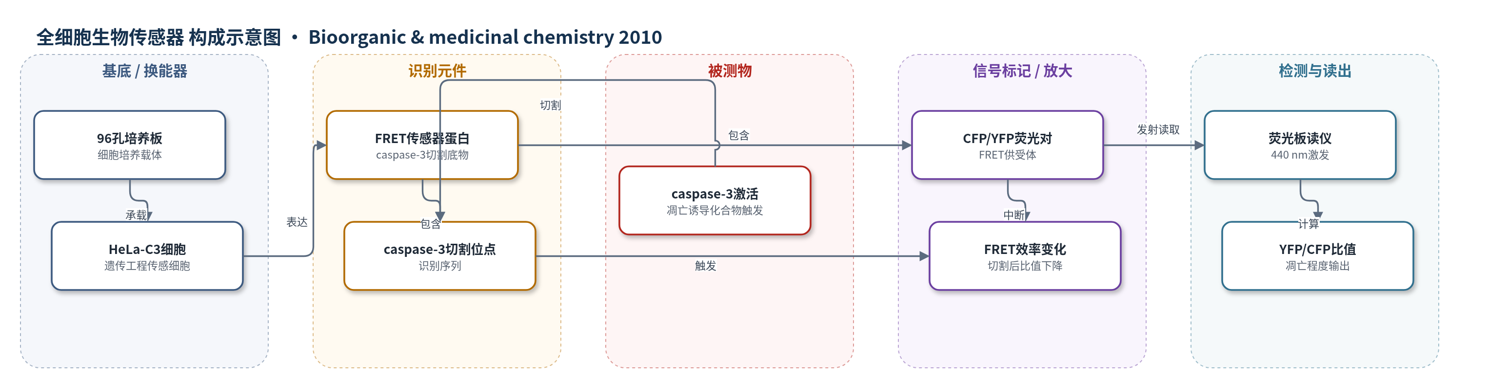
点击图片查看大图 · 依据论文自动绘制
传感器类型
全细胞生物传感器
检测对象
caspase-3 激活(caspase-3 activation)/凋亡程度(apoptosis),样品基质:HeLa-C3 细胞培养体系(96孔板细胞培养液)
检测原理
HeLa-C3细胞表达FRET型caspase-3传感器蛋白,该蛋白由CFP供体、YFP受体和caspase-3特异性切割序列组成。正常细胞中caspase-3未激活,传感器蛋白完整,CFP受激发后能量转移至YFP,YFP/CFP发射比值较高,通常约6–8。凋亡诱导化合物处理后,细胞内caspase-3被激活并切割传感器蛋白,使CFP与YFP空间距离增大,FRET效率下降,YFP发射降低而CFP相对升高,YFP/CFP比值降至3或以下。荧光板读仪在440 nm激发,分别测量486 nm CFP和535 nm YFP发射,通过比值变化非侵入地定量caspase-3激活程度和凋亡水平。该方法不依赖酶催化沉积或核酸放大,直接以FRET比值作为读出信号。
检测灵敏度
原文未报告LOD、线性范围、灵敏度斜率或R^2;仅报告阳性阈值:YFP/CFP emission ratio ≤ 3,正常细胞比值通常约6–8。
效应效果
19个化合物中8个在25 μM、72 h内使HeLa-C3细胞YFP/CFP比值降至≤3;化合物14在10 μM、72 h内仍阳性,提示其诱导凋亡最强。动力学显示化合物8–10在25 μM下24 h内降低比值,化合物2、5、14在48 h内降低,化合物6、11在72 h内降低。形态学见细胞收缩和脱落,Western blot证实化合物2、9、14处理36 h后PARP切割。MTT法IC50:化合物2为9.5±0.2 μM,化合物4为29.5±0.2 μM,化合物3为52.5±1.5 μM,化合物1为95.6±5.5 μM。作者认为paucinervin B抑制HeLa生长最强,具潜在抗癌价值。
传感器的构成
- 基底/培养载体:96孔细胞培养板,承载HeLa-C3细胞并提供检测微环境
- 换能器/传感细胞:遗传工程HeLa-C3细胞,表达FRET型caspase-3传感器蛋白,将酶活变化转换为荧光信号
- 识别元件:传感器蛋白中的caspase-3特异性切割位点,识别并响应caspase-3激活
- 信号标记物:CFP/YFP荧光蛋白对,作为FRET供体/受体,切割后FRET效率改变
- 信号读出:Perkin-Elmer Victor荧光板读仪,440 nm激发,486 nm测CFP、535 nm测YFP,输出YFP/CFP发射比值
中文摘要
从少叶藤黄(Garcinia paucinervis)叶片中分离得到4个新化合物paucinervins A–D(1–4)和15个已知化合物,并通过光谱证据解析新化合物结构。所有19个化合物均使用遗传工程HeLa-C3细胞评估其诱导凋亡作用;该细胞表达一种可检测caspase-3激活的荧光生物传感器。结果显示,8个化合物在25 μM浓度下72 h内可激活HeLa-C3细胞中的caspase-3。进一步用MTT法测定4个新化合物对HeLa细胞的IC50值,其中化合物2(paucinervin B)的IC50最低,为9.5 μM,其余三个新化合物分别为29.5、52.5和95.6 μM。结果表明,paucinervin B对HeLa细胞生长的抑制作用最强,可能具有开发为新型抗癌候选药物的潜力。
英文摘要
Four new compounds, paucinervins A-D (1-4), and 15 known ones were isolated from the leaves of Garcinia paucinervis. The structures of the new compounds were elucidated by spectroscopic evidences. All of the 19 compounds were evaluated for their apoptosis-inducing effects using HeLa-C3 cells which have been genetically engineered to possess a fluorescent biosensor capable of detecting caspase-3 activation. Eight of them were found to activate caspase-3 in HeLa-C3 cells within 72 h at the concentration of 25 microM. Moreover, the values of IC50 were measured for all four new compounds on HeLa cells using the MTT assay. Among them, compound 2 (paucinervin B) had the lowest IC50 value of 9.5 microM, while the other three new compounds had much higher IC50 values of 29.5, 52.5, and 95.6 microM, respectively. This result shows that paucinervin B has the strongest inhibitory effect against HeLa cell growth among these four newly identified paucinervins and it may have the potential to be developed into a new anticancer candidate.