组成图示
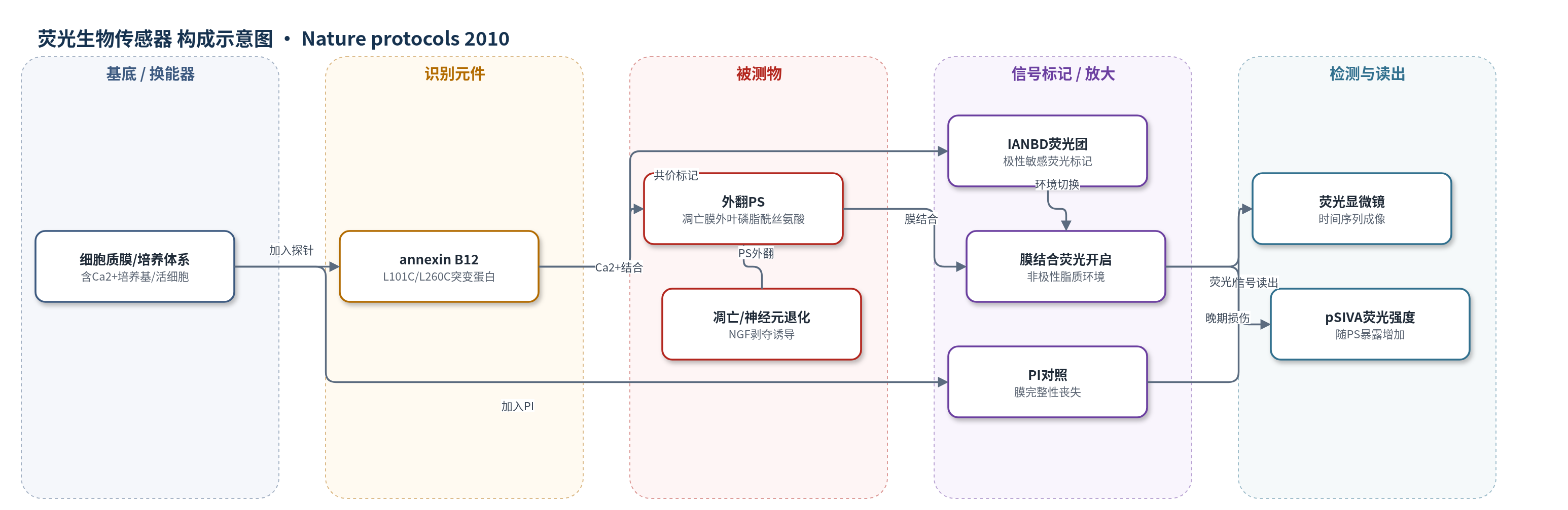
点击图片查看大图 · 依据论文自动绘制
传感器类型
荧光生物传感器
检测对象
凋亡细胞膜外翻的磷脂酰丝氨酸(phosphatidylserine, PS)、神经元退化/凋亡(neuronal degeneration/apoptosis);样品基质:原代背根神经节(DRG)神经元培养物、含Ca2+细胞培养基,亦可用于活体神经损伤组织。
检测原理
pSIVA由annexin B12(L101C/L260C)与极性敏感荧光团IANBD共价标记构成。在含Ca2+的培养体系中,annexin B12通过膜结合环以Ca2+依赖方式识别并结合凋亡细胞质膜外叶暴露的磷脂酰丝氨酸(PS)。未结合时,IANBD处于极性水相,荧光很弱;结合后,位于膜结合环的IANBD进入非极性脂质双分子层环境,荧光显著增强(约50倍)。因此,PS外翻面积或程度增加时,pSIVA荧光强度随之升高,形成与凋亡/神经元退化进程相关的荧光信号。荧光显微镜时间序列成像读取该信号,并可联合PI区分晚期膜完整性丧失。
检测灵敏度
原文未报告LOD、线性范围、灵敏度斜率或相关系数。
效应效果
pSIVA在溶液中荧光相对膜结合态约低50倍,可过量使用而不显著增加背景,无需洗涤即可连续活细胞成像。1 L大肠杆菌培养约获15 mg蛋白,纯度约99.9%,IANBD标记化学计量约2:1,标记蛋白避光保存数月稳定。在NGF剥夺诱导的原代DRG神经元中,pSIVA荧光最早约10 h出现,并在24-40 h内沿轴突逐渐增强,成像间隔约30 min;pSIVA工作浓度5-10 µg ml-1,PI 0.3 µg ml-1,持续存在10 µg ml-1未见明显毒性。与常规annexin流式/固定荧光显微镜单时间点检测相比,pSIVA可实时追踪PS外翻、凋亡与坏死区分及挽救过程。
传感器的构成
- 识别元件:annexin B12(L101C, L260C)突变蛋白,Ca2+依赖结合凋亡细胞膜外翻的磷脂酰丝氨酸(PS)
- 信号标记物:IANBD(N,N′-di-methyl-N(iodoacetyl)-N′-(7-nitrobenz-2-oxa-1,3-diazol-4-yl)ethyleneamine),共价连接于Cys101/Cys260,膜结合态进入非极性脂质环境后荧光增强
- 结合界面:细胞质膜外叶PS(phosphatidylserine)作为识别靶标与荧光环境切换界面,Ca2+介导annexin结合
- 辅助对照标记:propidium iodide(PI),标记膜完整性丧失的晚期凋亡或坏死细胞,与pSIVA联合区分死亡模式
- 读出平台:荧光显微镜/时间序列成像系统(Zeiss Axiovert 200、Cascade 1K相机、绿色荧光滤片excitation 487 nm/emission 525 nm),读取pSIVA荧光
中文摘要
非侵入性实时成像在监测凋亡和神经元退化方面受限于成像策略与现有探针的技术瓶颈。追踪细胞经历凋亡的过程可为理解启动细胞死亡的时间事件及挽救凋亡细胞提供重要信息。作者基于结构导向设计策略,构建了一种 annexin 基生物传感器,作为存活与凋亡的极性敏感指示剂(pSIVA),通过结合凋亡细胞膜上外翻的磷脂酰丝氨酸(PS)实现检测。pSIVA 仅在结合 PS 时发出荧光,在溶液中几乎不可检测。本文描述了 pSIVA 的设计、表达、纯化与标记方案,以及其在培养神经元退化时间序列成像中的应用;整个流程可在 2 周内完成。该方法的主要优势在于可灵活地与其他探针联用,在不扰动实验条件的前提下,实时研究凋亡与退化相关的细胞机制。
英文摘要
Applications for noninvasive real-time imaging of apoptosis and neuronal degeneration are hindered by technical limitations in imaging strategies and by existing probes. Monitoring the progression of a cell through apoptosis could provide valuable insight into the temporal events that initiate cell death as well as the potential for rescue of apoptotic cells. We engineered an annexin-based biosensor to function as a polarity-sensitive indicator for viability and apoptosis (known as pSIVA) by binding to externalized phosphatidylserine (PS) exposed on apoptotic cell membranes. Constructed from a structure-based design strategy, pSIVA fluoresces only when bound to PS and remains effectively undetectable in solution. In this paper, we describe protocols for the design, expression, purification and labeling of pSIVA as well as for its application in time-lapse imaging of degenerating neurons in culture; the entire protocol can be completed in 2 weeks. The primary advantage of this method is the flexibility to use pSIVA, in combination with other probes and without perturbing experimental conditions, to explore the cellular mechanisms involved in apoptosis and degeneration in real time.