组成图示
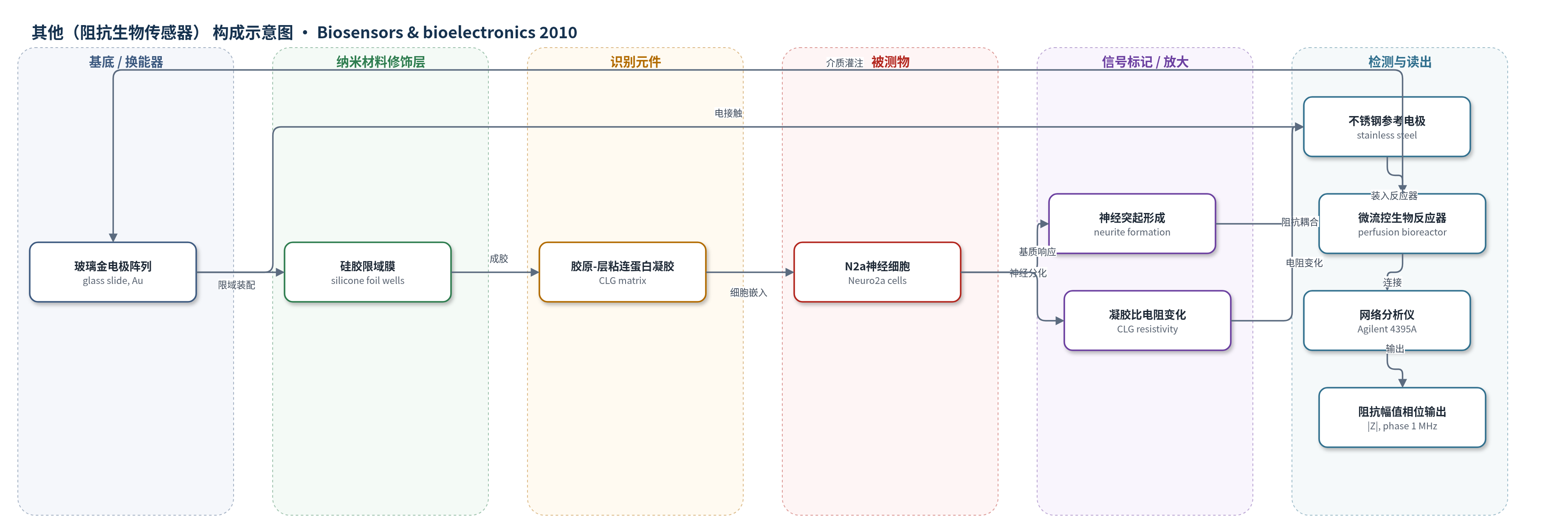
点击图片查看大图 · 依据论文自动绘制
传感器类型
其他(阻抗生物传感器)
检测对象
神经细胞分化状态(neuronal differentiation)与神经突起形成(neurite formation);样品基质:胶原-层粘连蛋白凝胶(CLG)、胶原凝胶(CG)等三维凝胶中的N2a神经母细胞瘤细胞(Neuro2a/N2a cells),无血清DMEM。
检测原理
该传感器以交流阻抗谱作为换能机制。N2a神经母细胞瘤细胞被嵌入胶原-层粘连蛋白凝胶(CLG)中,在层粘连蛋白和无血清DMEM作用下发生神经分化并形成神经突起。细胞膜可等效为电阻-电容元件,突起使细胞膜表面积和细胞-基质界面增加,改变局部介电与导电性质;同时凝胶中离子迁移受限,胶原-层粘连蛋白基质的比电阻也随时间变化。传感器在100 kHz–50 MHz范围内施加交流电场,重点在1 MHz读取阻抗幅值和相位。随着分化程度增加,阻抗幅值上升,相位基本稳定。信号由金电极阵列与多孔不锈钢参考电极采集,经网络分析仪读出,无需外源标记或酶放大。
检测灵敏度
未报告LOD、线性范围、灵敏度斜率或相关系数。
效应效果
五种凝胶基质中,仅BME和CLG支持N2a细胞分化,CLG促进较长神经突起;db-cAMP仅轻微增强。细胞密度≤2×10^6 cells/mL分化较高,>2×10^6 cells/mL部分丢失;MTT吸光度在4×10^6 cells/mL最大,>4×10^6 cells/mL活力下降。12 h阻抗监测中,1 MHz下裸CG前2 h约7%变化来自热平衡,之后稳定;CLG中阻抗幅值随分化增加,初始最多降低约2%。温度稳定性0.1 K,四电极并联可识别气泡或填充不良,交流电流和介质流动对细胞无显著影响。作者认为该模型可用于发育神经生物学药物筛选。
传感器的构成
- 基底/测量电极:玻璃片(glass slide)上溅射16个金电极(Au,300 nm±20 nm,铬底层),用于施加交流电场并采集阻抗信号。
- 限域/成型层:1 mm厚硅胶膜(silicone foil)带Ø5 mm圆形孔,限定凝胶膜形状与厚度,保证有效测量面积。
- 识别/响应基质:胶原-层粘连蛋白凝胶(CLG)或胶原凝胶(CG)等三维基质,层粘连蛋白介导细胞黏附与神经分化。
- 识别/响应细胞:N2a神经母细胞瘤细胞(Neuro2a/N2a cells),嵌入凝胶中,其细胞膜与神经突起作为内源响应元件。
- 参考/接地电极:多孔不锈钢薄板(stainless steel sheet),作为接地参考电极,与金电极夹持凝胶膜形成电接触。
- 微流控灌注层:玻璃/硅胶微流道(micro-fluidic channels)与不锈钢生物反应器,四个入口/出口,持续灌注无血清DMEM及刺激物。
- 信号读出层:网络分析仪(Agilent 4395A)阻抗模式,100 kHz–50 MHz,1 MHz监测阻抗幅值与相位。
中文摘要
神经细胞分化是中枢神经系统发育的关键过程,对再生医学等领域具有重要意义。本研究报道了一种用于研究凝胶基质中神经细胞分化与信号过程的新型阻抗生物传感器。作者将Neuro2a(N2a)神经母细胞瘤细胞固定于不同三维凝胶基质中,比较其分化程度,并分析细胞数量及分化因子的影响。结果表明,胶原-层粘连蛋白混合凝胶在无血清培养基中可诱导神经分化,而胶原、琼脂糖或细菌琼脂基质不能支持分化;层粘连蛋白是三维胶原基质中分化的关键因素,db-cAMP仅轻微促进神经突起形成。胶原-层粘连蛋白凝胶因此可作为发育神经生物学药物筛选的三维模型。优化固定过程后,作者利用新型阻抗传感器和电化学阻抗谱技术,通过监测介电与导电性质变化在线跟踪分化过程。结果显示,分化过程中阻抗幅值增加,主要归因于分化细胞及神经突起形成,同时伴随裸胶原-层粘连蛋白凝胶比电阻的增加。
英文摘要
The differentiation of neural cells is an important process during the development of the central nervous system. Studies on the mechanisms of the differentiation process is of special importance, e.g. in the field of regenerative medicine. In this contribution the cellular differentiation of gel matrix embedded neuronal cells was studied. The three-dimensional organization of neuronal cells represents a new cellular model system closer to the physiology than conventional two-dimensional cell cultures. Neuro2a (N2a) neuroblastoma cells were immobilized in different gel matrices and the grade of differentiation was compared. Furthermore, the impact of the cell number and selected differentiation factors were analyzed. Experimental results revealed that gel matrices based on collagen-laminin mixtures in contact with serum free medium enable neural differentiation. Therefore, collagen-laminin gels appear as a suitable three-dimensional model for drug screening in developmental neurobiology. Following optimization of the immobilization process, a novel impedimetric sensor and electrical impedance spectroscopy technique was applied to on-line monitor the differentiation process by means of changes in the dielectric and conductive properties. Experimental results showed an increase in the impedance magnitude that can be mainly attributed to differentiating cells accompanied by an increase in the specific resistivity of the bare gel mixture.