组成图示
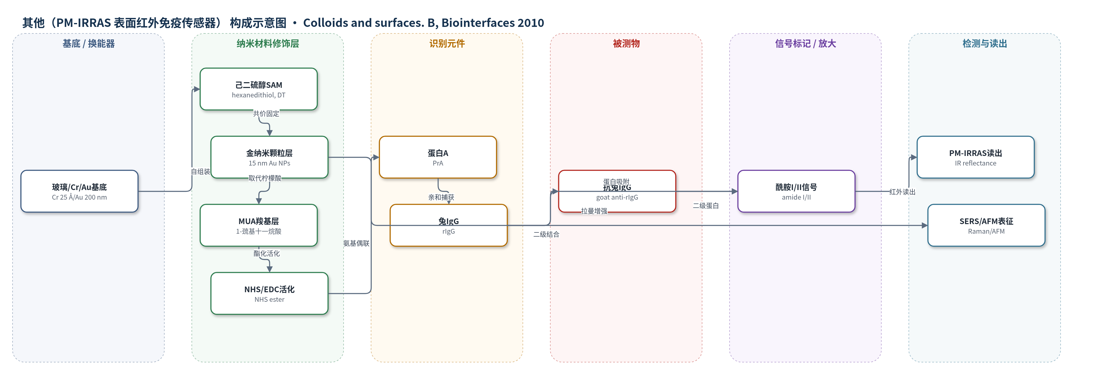
点击图片查看大图 · 依据论文自动绘制
传感器类型
其他(PM-IRRAS 表面红外免疫传感器)
检测对象
兔免疫球蛋白(rabbit IgG, rIgG)、山羊抗兔 IgG(goat anti-rIgG);样品基质:PBS 缓冲液、山羊血清
检测原理
该传感器以金表面为换能基底,通过己二硫醇自组装层将 15 nm 金纳米颗粒共价固定,形成三维界面。MUA 取代柠檬酸后,NHS/EDC 活化羧基,使蛋白 A 共价固定;PrA 通过 Fc 亲和结合兔 IgG,山羊抗兔 IgG 再结合 rIgG 恒定区。蛋白吸附使界面酰胺 I(约 1655 cm−1)和酰胺 II(约 1550 cm−1)红外吸收增强,PM-IRRAS 通过 p/s 偏振差分反射率定量吸附量。金纳米颗粒同时提供 SERS 增强,用于验证纳米颗粒和界面组装。三维结构增加比表面积和抗体固定量,使 rIgG 密度提高约 4 倍,但二级抗体结合比略降,说明可及性仍保持。未来可结合 SPR 利用纳米颗粒等离子效应进一步放大信号。
检测灵敏度
原文未报告 LOD、线性范围、灵敏度斜率或相关系数。
效应效果
BSA 封闭后山羊血清处理未改变酰胺带面积,表明非特异吸附被抑制。与平面金相比,三维 Au NPs 表面 PrA 固定量几乎加倍,rIgG 固定量提高约 4 倍;anti-rIgG/rIgG 比值由平面的 2–3 降至三维的约 2,可及性略降但仍较高。AFM 显示室温 1 h 沉积形成约 16–18 nm 单层,覆盖约 35%,3 h 出现约 30 nm 聚集体;XPS 表明覆盖仍较低。DT 在乙醇 4 °C 3 h 下形成伸展单分子层(S/Au=0.010,Sb/Sf=1.2),优于水相 12 h 多层(S/Au=0.049)。作者认为该策略可构建高密度抗体阵列,并预期结合 SPR 可提升约一个数量级灵敏度。
传感器的构成
- 基底/换能器:玻璃基底上依次溅射 25 Å Cr 和 200 nm Au,提供金表面与光学/表面分析基底
- 二硫醇自组装层:己二硫醇(hexanedithiol, DT)在乙醇中 4 °C 浸泡 3 h 形成伸展单分子层,一端 Au–S 键合,一端保留自由 –SH
- 金纳米颗粒层:15 nm 柠檬酸稳定球形 Au NPs(Turkevich 法)通过自由 –SH 形成 S–Au 共价键,形成三维表面
- 羧基硫醇修饰层:1-巯基十一烷酸(MUA)取代 Au NPs 表面柠檬酸,提供 –COOH 用于蛋白偶联
- 活化层:NHS(60 mM)和 EDC(30 mM)将 –COOH 转化为 NHS 酯,用于与蛋白氨基共价偶联
- 识别元件:重组蛋白 A(PrA)通过氨基与 NHS 酯偶联,用于亲和捕获 IgG 的 Fc 片段
- 封闭剂:牛血清白蛋白(BSA)封闭非特异结合位点,降低山羊血清蛋白非特异吸附
- 模型抗体/检测抗体:兔 IgG(rIgG)固定于 PrA 上作为模型捕获抗体;山羊抗兔 IgG(anti-rIgG)用于检测 rIgG 可及性
中文摘要
本文报道了在平面金表面可控固定金纳米颗粒并构建三维生物传感器的方法。首先用己二硫醇(hexanedithiol, DT)功能化金表面,其两个硫醇端基使一端接枝到金上,另一端保留自由硫醇用于连接纳米颗粒。通过改变溶剂、温度和浸泡时间优化接枝条件,并用 PM-IRRAS 和 XPS 监测。高分辨 XPS 硫峰区分结合硫与自由硫,从而确定使 DT 以伸展构型接枝的最佳条件。随后将 15 nm 球形金纳米颗粒固定到表面,用 SERS 和 AFM 证实。所得金层用于构建三维免疫传感器,依次接枝蛋白 A(PrA)、兔 IgG(rIgG)和 BSA,各步用 PM-IRRAS 表征并与平面金比较。尽管纳米颗粒小且密度低,PrA 固定量几乎加倍,rIgG 固定量比平面高约 4 倍,但二级 IgG 识别显示可及性略降。该工作证明纳米颗粒阵列固定分子受体的可行性,并强调控制各步条件对优化受体数量至关重要。
英文摘要
This paper describes a controlled way to immobilize gold nanoparticles on planar gold surfaces and the use of the resulting 3D platform to build up a 3D biosensor. The surface was first functionalized by grafting hexanedithiol, this molecule has 2 thiol end groups, which enables its chemical grafting to planar gold while retaining a free thiol group to attach nanoparticles. This step was optimized by varying experimental parameters such as solvent, temperature and immersion time. The grafting was monitored by polarization modulation infrared reflection absorption spectroscopy (PM-IRRAS) and X-ray photoelectron spectroscopy (XPS). The high resolution XPS sulfur peak made clear the existence of two contributions, S bound to gold and free S, thus led us to determine the optimal conditions to graft hexanedithiol in an extended conformation. 15 nm spherical gold nanoparticles were then immobilized on the resulting surface and their presence was evidenced by surface enhanced Raman spectroscopy (SERS) and atomic force microscopy (AFM). The resulting gold layer was used to build up a 3D biosensor by grafting protein A (PrA), rabbit immunoglobulin (rIgG), and bovine serum albumin (BSA), respectively. Each step was characterized by PM-IRRAS then compared to the results on planar gold surface. Despite the small size of particles and their rather low density on the planar surface, the amount of immobilized proteins, starting from PrA, was almost doubled. The amount of rIgG fixed on the 3D layer was also significantly increased ( approximately 4 times higher than on planar surfaces), however accompanied by a slight decrease of their accessibility, checked by assaying the recognition of a secondary IgG. This work demonstrates the feasibility and interest of building arrays of nanoparticles to immobilize molecular receptors; it also shows that controlling the conditions of elaboration of the biosensor at each step is determining for optimizing the number of molecular receptors.