组成图示
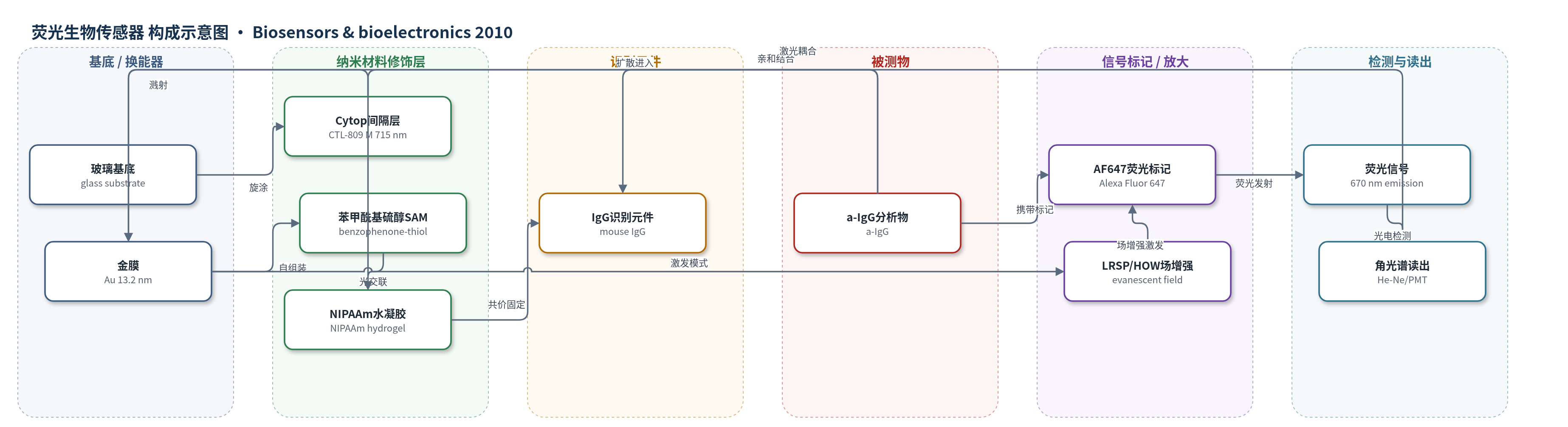
点击图片查看大图 · 依据论文自动绘制
传感器类型
荧光生物传感器
检测对象
山羊抗小鼠免疫球蛋白G(goat anti-mouse IgG, a-IgG,Alexa Fluor 647标记),样品基质为PBS/Tween 20(PBST)加标液
检测原理
样品中Alexa Fluor 647标记的山羊抗小鼠IgG(a-IgG-AF647)经流池扩散进入羧基化NIPAAm水凝胶,与凝胶内共价固定的小鼠IgG发生亲和结合。金膜/Cytop结构支持长程表面等离子体(LRSP)或水凝胶光波导(HOW)模式,共振激发的倏逝电磁场可穿透数百纳米至微米级水凝胶,并在界面附近形成强场增强。该增强场激发结合态AF647发射670 nm荧光,荧光强度正比于探测场内捕获的a-IgG数量。低浓度下结合受扩散限制,荧光随时间线性上升,斜率与浓度成正比;通过匹配水凝胶厚度、扩散深度与倏逝场轮廓,使结合事件位于高场区,从而实现飞摩尔级检测。
检测灵敏度
LOD: below 20 fM;线性范围: 6.7 fM–6.7 pM(低浓度下荧光随时间线性增加);LRSP灵敏度斜率: dF/(dt·a0)=0.34 cps min^-1 fM^-1;HOW灵敏度斜率: dF/(dt·a0)=0.59 cps min^-1 fM^-1
效应效果
该传感器在模型免疫分析中实现低于20 fM的检出限。HOW模式在1440 nm水凝胶中的荧光信号比LRSP在130 nm水凝胶中高1.8倍;LRSP基线噪声为55 cps,最大斜率0.34 cps min^-1 fM^-1,HOW基线噪声107 cps,斜率0.59 cps min^-1 fM^-1。实验误差棒来自三片独立制备芯片的重复测量,但未给出RSD、稳定性、抗干扰、实际样品回收率或与ELISA/HPLC/qPCR的对比。作者认为大容量三维水凝胶结合基质尤其适用于低亲和识别元件或快速扩散小分子检测,并展望纳米结构、响应型和分子印迹水凝胶。
传感器的构成
- 基底:玻璃基底(glass substrate),承载功能层并用于棱镜光学耦合
- 光学间隔层:Cytop氟聚合物(CTL-809 M,9 wt.% in CT-solv 180,约715 nm),与金膜共同支持长程表面等离子体(LRSP)
- 金属换能层:金薄膜(Au,约13.2 nm),激发LRSP/HOW倏逝场并增强近场
- 自组装单分子层:苯甲酰基硫醇(benzophenone-thiol,约2 nm),通过苯甲酰基光交联连接水凝胶
- 三维结合基质:羧基化聚(N-异丙基丙烯酰胺)三元共聚物水凝胶(NIPAAm/甲基丙烯酸/4-甲基丙烯酰基苯甲酮,溶胀厚度可达微米级),容纳抗体并允许分析物扩散
- 识别元件:小鼠免疫球蛋白G(mouse IgG),经TFPS/EDC活化羧基共价固定于水凝胶中,捕获目标a-IgG
- 封闭剂:乙醇胺(ethanolamine,1 M,pH 8.5),封闭未反应TFPS酯基
- 信号标记物:Alexa Fluor 647标记的山羊抗小鼠IgG(a-IgG-AF647),结合后产生荧光信号
- 样品流路:PDMS垫片与透明石英片构成的流池(深度0.3 mm),使PBST样品流过传感器表面
中文摘要
本文研究了一种基于倏逝波亲和生物传感器与大容量三维结合基质的实现,用于分子分析物的超灵敏检测。实验中,将厚度可达微米级的高度溶胀羧基化聚(N-异丙基丙烯酰胺)(NIPAAm)水凝胶接枝到传感器表面,并用抗体识别元件功能化,用于检测液体样品中的目标分子。分子结合事件通过长程表面等离子体(LRSP)和水凝胶光波导(HOW)场增强荧光光谱进行检测。这些新方法可探测扩展的三维生物界面,倏逝场可从传感器表面延伸至数微米。LRSP 和 HOW 模式的共振激发提供强电磁场增强,直接转化为与荧光标记分子结合相关的增强荧光信号。实验观察得到目标分子在水凝胶中质量传递和亲和结合数值模拟的支持。通过优化水凝胶厚度和探测倏逝波轮廓,实现了低飞摩尔级检出限。
英文摘要
An implementation of evanescent wave affinity biosensor with a large-capacity three-dimensional binding matrix for ultra-sensitive detection of molecular analytes is investigated. In the experimental part of the work, highly swollen carboxylated poly(N-isopropylacryamide) (NIPAAm) hydrogel with up to micrometer thickness was grafted to a sensor surface, functionalized with antibody recognition elements and employed for immunoassay-based detection of target molecules contained in a liquid sample. Molecular binding events were detected by long range surface plasmon (LRSP) and hydrogel optical waveguide (HOW) field-enhanced fluorescence spectroscopy. These novel methods allowed probing an extended three-dimensional biointerface with an evanescent field reaching up to several micrometers from the sensor surface. The resonant excitation of LRSP and HOW modes provided strong enhancement of intensity of electromagnetic field that is directly translated into an increased fluorescence signal associated with the binding of fluorophore-labeled molecules. Experimental observations were supported by numerical simulations of mass transfer and affinity binding of target molecules in the hydrogel. Through the optimization of the hydrogel thickness and profile of the probing evanescent wave, low femtomolar limit of detection was achieved.