组成图示
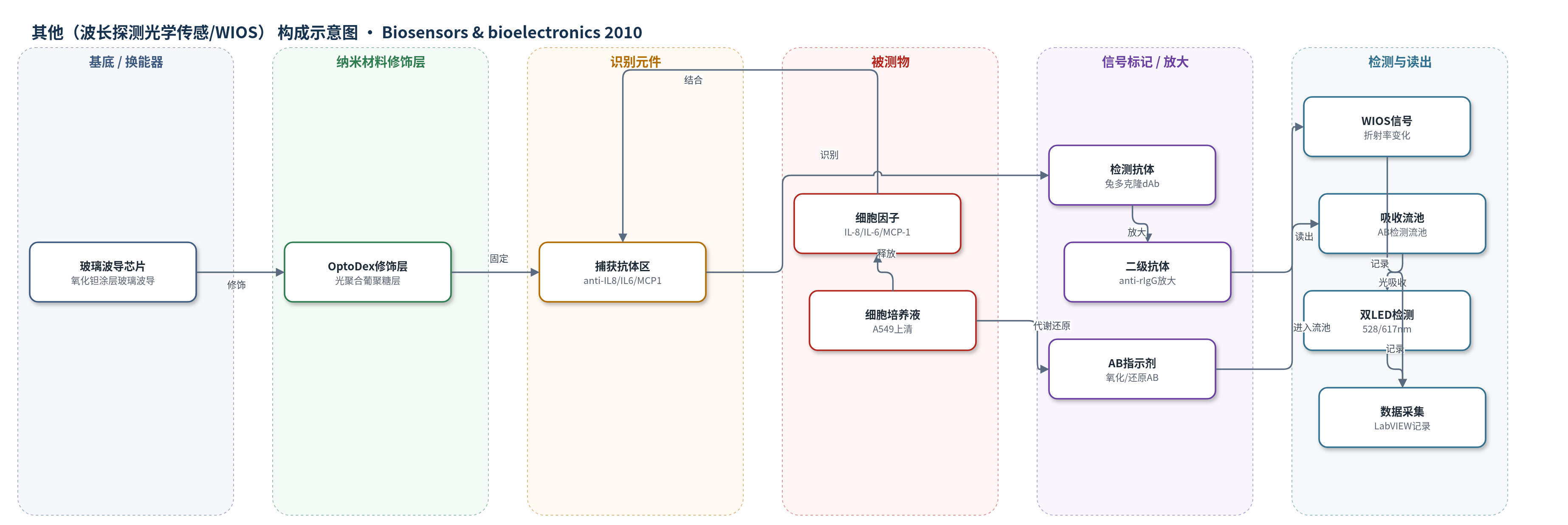
点击图片查看大图 · 依据论文自动绘制
传感器类型
其他(波长探测光学传感/WIOS)
检测对象
白细胞介素8(IL-8)、白细胞介素6(IL-6)、单核细胞趋化蛋白-1(MCP-1);辅助检测细胞增殖(AlamarBlue, AB);样品基质:A549细胞培养上清/培养基
检测原理
WIOS基于波导光栅倏逝场探测芯片表面折射率变化。OptoDex层上固定捕获抗体,细胞培养液中的IL-8、IL-6或MCP-1与对应捕获抗体结合;随后加入检测抗体和抗兔IgG二级抗体,形成夹心免疫复合物,使表面质量和折射率增加,引起共振波长偏移。WIOS通过扫描共振波长读出信号,信号随细胞因子浓度升高而增大。芯片多区可分别检测三种细胞因子,并用参考区扣除漂移。AB增殖检测中,细胞代谢将氧化态AB(蓝色,600 nm)还原为还原态(红色,570 nm),在线吸收流池用双LED测量528/617 nm信号比,反映细胞活力和增殖状态。
检测灵敏度
灵敏度: 1–100 ng/mL(依细胞因子而异);IL-8: 1 ng/mL;IL-6: 10 ng/mL;MCP-1: 100 ng/mL;WIOS表面灵敏度: ∼1 ng/cm²;校准范围: IL-8 0–10 ng/mL、IL-6 0–50 ng/mL、MCP-1 0–1000 ng/mL
效应效果
三种细胞因子之间未观察到交叉反应,混合样品响应与单独检测相当。表面再生每周期导致约10% WIOS信号损失,可在连续实验中校正;完整免疫分析(含再生)约90 min。AB LED比值测量误差为2%;AB试剂接触24 h会引起细胞收缩脱落,因此仅在测量前6 h加入。rhTNF-α诱导细胞释放的IL-8显著高于对照,而金纳米颗粒未引起三种细胞因子释放,但48 h后影响细胞增殖。作者认为该平台通过封闭流体通道降低污染,将分析时间从1–2天缩短至2 h,可实时、多路评估纳米颗粒和化学物质的细胞毒性。
传感器的构成
- 基底/换能器:氧化钽涂层玻璃波导芯片(Tantalum oxide-coated glass waveguide chips),含三列八个独立波导光栅垫,通过倏逝场探测表面折射率变化
- 修饰层:OptoDex®光聚合葡聚糖层(photopolymerizable dextran layer),用于共价固定抗体并抑制非特异吸附
- 识别元件:捕获抗体(capture antibodies),包括anti-IL-8(JK8-1)、anti-IL-6(MQ2-13A5)、anti-MCP-1(2H5),分别印在独立测量区并经UV固定
- 检测抗体:兔多克隆检测抗体(dAb,anti-IL8/anti-IL6/anti-MCP1混合物,20 µg/mL),识别已捕获的细胞因子
- 信号放大元件:兔特异性IgG二级抗体(anti-rIgG,Ab2,50 µg/mL),增加免疫复合物质量以放大WIOS响应
- 流体/流池:PDMS垫片分隔的三个独立流体通道及Tygon管、注射泵、蠕动泵、六通阀,用于细胞培养液和试剂的原位输送
- 吸收测量单元:FIAlab吸收流池(FIA-Z-SMA-ULT,光程20 mm,体积120 µL)与双LED(528/617 nm)光电二极管,用于AB细胞增殖检测
中文摘要
开发了一种分析检测平台,用于评估暴露于生长因子或纳米颗粒等外源物质的细胞培养物所诱导的毒性。将生物传感检测装置连接至细胞培养瓶,可实时分析细胞培养基成分。该分析基于通过固相免疫分析定量细胞释放到培养基中的炎症细胞因子,并使用波长探测光学传感(WIOS)仪器进行检测。用于原位测量的流体系统可实时检测细胞因子,灵敏度因细胞因子而异,为1–100 ng/mL。此外,集成在线光学吸收测量单元,并结合标准AB细胞增殖试验,可提供培养体系中细胞活力信息。细胞培养瓶、光学生物传感器和吸收测量单元之间的流体连接可同时定量最多三种细胞因子(白细胞介素8、白细胞介素6和单核细胞趋化蛋白),评估细胞增殖,从而区分未诱导细胞与暴露于生长因子(肿瘤坏死因子α)或纳米颗粒等外源物质的细胞。该分析工具在体外评估纳米颗粒及其他化学物质细胞毒性方面具有较高潜力。
英文摘要
An analytical detection platform was developed to evaluate the induced toxicity in cell cultures exposed to foreign agents like growth factors or nanoparticles. Connecting a biosensing detection device to the cell culture flasks allows analyzing the composition of cell medium in real-time. The analysis relies on the quantification of inflammatory cytokines released by cells into the cell culture medium, by means of solid-phase immunoassays analyzed with the wavelength interrogated optical sensing (WIOS) instrument. A fluidic system for in situ measurements allows detecting cytokines in real-time, with a sensitivity of 1-100 ng/mL depending on the cytokine. In addition, integration of an in-line optical absorbance measurement unit, in combination with the standard AB cell proliferation assay, provides information on the cell viability in the culture. Fluidic connections between the cell culture flasks, the optical biosensor and the absorbance measurement unit simultaneously allow quantifying up to three cytokines (interleukin 8, interleukin 6 and the monocyte chemotactic protein), assessing cellular proliferation, and thus discriminating between naïve cells and cells exposed to foreign agents such as growth factors (tumor necrosis factor alpha) or nanoparticles. This analytical tool presents a high potential for assessing the cytotoxicity of nanoparticles and other chemicals in vitro.