组成图示
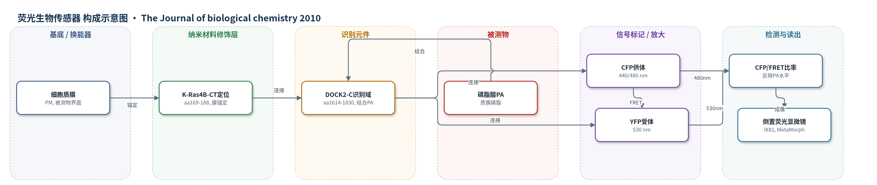
点击图片查看大图 · 依据论文自动绘制
传感器类型
荧光生物传感器
检测对象
磷脂酸(phosphatidic acid, PA);样品基质:活细胞质膜(COS7、HeLa、NIH3T3、MDCK 细胞培养物)
检测原理
Pii-DK 将 CFP 与人 DOCK2 C 端 PA 结合域、YFP 串联,并由 K-Ras4B-CT 锚定于质膜。当质膜 PA 浓度升高时,DOCK2-C 结合 PA,引起探针构象或荧光蛋白取向/距离变化,使 CFP 向 YFP 的 FRET 效率降低。用 440 nm 激发 CFP,同时采集 480 nm CFP 发射与 530 nm FRET 发射,计算 CFP/FRET 比率;PA 越高,FRET 越弱,CFP/FRET 越高。该法为比率型荧光读出,可抵消表达量与光漂白影响,实时反映活细胞质膜 PA 的时空变化。
检测灵敏度
原文未报告LOD、线性范围、灵敏度斜率或R^2。
效应效果
Pii-DK 对 PA 特异:Pii-DK-9A 与截短 SOS-PH 对照反应弱,PBS 仅可忽略变化;苯胂氧降低 PI(4,5)P2 但不影响 Pii-DK,特布他林升高 CFP/FRET。0.5% n-BtOH 与 750 nM FIPI 抑制 PLD 后,EGF 诱导的 PA 和 Ras 激活下降。EGF 50 ng/ml 使 COS7/HeLa PA 升高,NIH3T3 较弱,MDCK 不升高;ATP 可诱导短暂 PA。传感器揭示细胞系间及细胞内基础 PA 异质性,基础 PA 与诱导增量负相关;PA 在细胞自由边缘高于细胞-细胞接触区,与 Ras 激活分布一致。
传感器的构成
- 换能器/荧光对:CFP-YFP FRET 对,CFP 为供体、YFP 为受体,PA 结合改变 FRET 效率
- 供体荧光蛋白:CFP(cyan fluorescent protein,aa1–237),440 nm 激发/480 nm 发射
- 连接肽:(Glu-Ala-Ala-Ala-Arg)6 与 (EAAAAR)3-GG-(EAAAAR)3,连接结构域并允许构象变化
- 识别元件:人 DOCK2 C 端 PA 结合域(DOCK2-C,aa1614–1830),特异性结合 PA;Pii-DK-9A 为 9 个关键氨基酸 Ala 突变负对照
- 受体荧光蛋白:YFP(yellow fluorescent protein,aa1–237),530 nm 发射
- 膜定位层:K-Ras4B C 端(K-Ras4B-CT,aa169–188),法尼基化/膜靶向序列,将 Pii 锚定于质膜
- 表达平台:质粒转染活细胞(COS7、HeLa、NIH3T3、MDCK),在质膜表达 Pii
中文摘要
磷脂酸(PA)是质膜中的主要磷脂之一。尽管已有报道表明 PA 在细胞存活和形态中发挥关键作用,但在活细胞中 PA 何时、何地产生仍不清楚。基于 Förster 共振能量转移(FRET)原理,作者构建了 PA 生物传感器并命名为 Pii(phosphatidic acid indicator)。该传感器将 DOCK2 的脂质结合域夹在青色荧光蛋白(CFP)与黄色荧光蛋白(YFP)之间,并带有 K-Ras 的质膜靶向序列。加入合成 PA,或在质膜激活磷脂酶 D(PLD)或二酰甘油激酶(DGK),均改变表达 Pii 的细胞中 FRET 水平,证明 Pii 对 PA 的响应。该传感器还检测到不同细胞系之间以及同一细胞系内 PA 含量的差异。有趣的是,生长因子诱导的 PA 增量与刺激前基础 PA 含量呈负相关,提示质膜 PA 浓度存在上限阈值。传感器还揭示细胞内 PA 分布不均:基础水平和生长因子诱导的 PA 积累在细胞自由边缘高于细胞-细胞接触区。PA 增加不足可能解释细胞-细胞接触区 Ras 激活无效。总之,PA 生物传感器 Pii 是研究单细胞及不同细胞间 PA 含量与分布异质性的通用工具。
英文摘要
Phosphatidic acid (PA) is one of the major phospholipids in the plasma membrane. Although it has been reported that PA plays key roles in cell survival and morphology, it remains unknown when and where PA is produced in the living cell. Based on the principle of Förster resonance energy transfer (FRET), we generated PA biosensor, and named Pii (phosphatidic acid indicator). In these biosensors, the lipid-binding domain of DOCK2 is sandwiched with the cyan fluorescent protein and yellow fluorescent protein and is tagged with the plasma membrane-targeting sequence of K-Ras. The addition of synthetic PA, or the activation of phospholipase D or diacylglycerol kinase at the plasma membrane, changed the level of FRET in Pii-expressing cells, demonstrating the response of Pii to PA. The biosensor also detected divergent PA content among various cell lines as well as within one cell line. Interestingly, the growth factor-induced increment in PA content correlated negatively with the basal PA content before stimulation, suggesting the presence of an upper threshold in the PA concentration at the plasma membrane. The biosensor also revealed uneven PA distribution within the cell, i.e. the basal level and growth factor-induced accumulation of PA was higher at the cell-free edges than at the cell-cell contact region. An insufficient increase in PA may account for ineffective Ras activation at areas of cell-cell contact. In conclusion, the PA biosensor Pii is a versatile tool for examining heterogeneity in the content and distribution of PA in single cells as well as among different cells.