组成图示
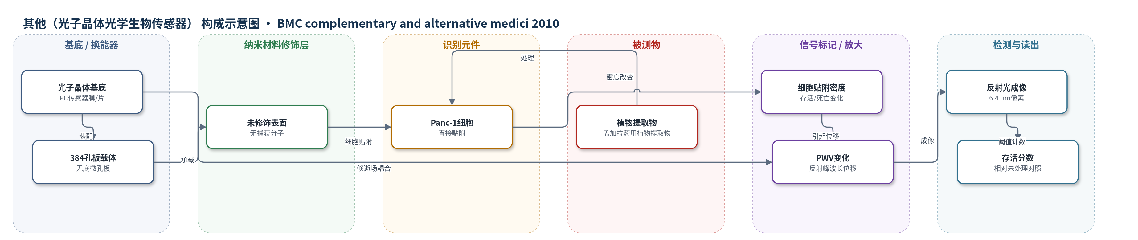
点击图片查看大图 · 依据论文自动绘制
传感器类型
其他(光子晶体光学生物传感器)
检测对象
孟加拉药用植物粗提取物(Bangladeshi medicinal plant crude extracts)诱导的Panc-1细胞贴附/存活变化;样品基质:细胞培养液(DMEM/IMDM,乙醇≤0.5% v/v)
检测原理
PC生物传感器表面存在倏逝场,当Panc-1细胞贴附于传感器表面时,局部质量/折射率变化使反射光峰值波长(PWV)发生位移。实验先采集无细胞基线图像,再采集细胞贴附及提取物处理后图像,通过图像对齐和基线减法得到PWV位移图。植物提取物若诱导细胞死亡或抑制增殖,贴附的活细胞密度下降,PWV位移减小;若促进增殖则位移增大。系统按6.4 μm×6.4 μm像素阈值计数,将PWV位移转换为相对未处理对照的细胞存活分数。该过程无标记、无需染色,信号随提取物细胞毒性增强而降低。
检测灵敏度
未报告LOD、线性范围、灵敏度斜率、R^2。
效应效果
PC筛选56种提取物,100 μg/mL处理24 h后22种使Panc-1存活率降>50%,9种>80%死亡;MTT确认3种在三种胰腺癌细胞中>70%死亡。IC50范围:P. punctata 20.3–31.4、A. sessilis 13.08–34.9、A. chittagonga 42.8–49.8 μg/mL;仅A. chittagonga对Hs68低毒(IC50>100 μg/mL)。P. punctata诱导caspase-3活性接近staurosporine。PC约500细胞/孔,低于MTT 2500–3000,与MTT一致,适合高通量筛选。
传感器的构成
- 换能基底:光子晶体(PC)传感器膜/片,利用倏逝场和反射光峰值波长(PWV)变化实现无标记检测
- 微孔板载体:无底384孔板,承载PC传感器并兼容标准微孔板加样与成像
- 识别/结合界面:未修饰PC表面,Panc-1细胞直接贴附(无特异性捕获分子),用于细胞贴附密度检测
- 信号响应层:贴附的Panc-1细胞,其存活/增殖改变引起局部贴附密度变化,进而改变PWV
- 信号读出层:反射光成像系统,按6.4 μm×6.4 μm像素记录PWV图像,通过基线减法获得细胞贴附变化
中文摘要
胰腺癌死亡率高且缺乏有效化疗药物,天然产物是抗肿瘤药物发现的重要来源。本研究对孟加拉传统药用植物库中44种植物的56种粗提取物进行抗胰腺癌活性筛选。首先采用无标记光子晶体(PC)生物传感器细胞贴附法检测提取物对Panc-1胰腺腺癌细胞的细胞毒性,通过反射光峰值波长(PWV)图像量化细胞存活变化;随后对筛选出的强毒性提取物采用MTT增殖法在Mia-Paca2、Capan-1两种胰腺癌细胞及Hs68成纤维细胞中验证,并用caspase-3比色法检测凋亡。结果显示,Petunia punctata、Alternanthera sessilis和Amoora chittagonga对三种胰腺癌细胞具有显著细胞毒性,IC50分别为20.3–31.4、13.08–34.9和42.8–49.8 μg/mL;P. punctata处理增加caspase-3活性,提示其细胞毒性由凋亡介导。仅A. chittagonga对Hs68成纤维细胞毒性较低,IC50>100 μg/mL。该工作为后续活性成分鉴定和机制研究提供候选植物提取物。
英文摘要
BACKGROUND: There has been a long standing interest in the identification of medicinal plants and derived natural products for developing cancer therapeutics. Our study focuses upon pancreatic cancer, due to its high mortality rate, that is attributed in part to the lack of an effective chemotherapeutic agent. Previous reports on the use of medicinal plant extracts either alone or alongside conventional anticancer agents in the treatment of this cancer have shown promising results. This work aims to investigate the therapeutic properties of a library of medicinal plants from Bangladesh.
METHODS: 56 extracts of 44 unique medicinal plants were studied. The extracts were screened for cytotoxicity against the pancreatic adenocarcinoma cell line Panc-1, using a label-free biosensor assay. The top cytotoxic extracts identified in this screen were tested on two additional pancreatic cancer cell lines (Mia-Paca2 and Capan-1) and a fibroblast cell line (Hs68) using an MTT proliferation assay. Finally, one of the most promising extracts was studied using a caspase-3 colorimetric assay to identify induction of apoptosis.
RESULTS: Crude extracts of Petunia punctata, Alternanthera sessilis, and Amoora chittagonga showed cytotoxicity to three cancer cell lines with IC50 values ranging between 20.3 - 31.4 μg/mL, 13.08 - 34.9 μg/mL, and 42.8 - 49.8 μg/mL, respectively. Furthermore, treatment of Panc-1 cells with Petunia punctata was shown to increase caspase-3 activity, indicating that the observed cytotoxicity was mediated via apoptosis. Only Amoora chittagonga showed low cytotoxicity to fibroblast cells with an IC50 value > 100 μg/mL.
CONCLUSION: Based upon the initial screening work reported here, further studies aimed at the identification of active components of these three extracts and the elucidation of their mechanisms as cancer therapeutics are warranted.