组成图示
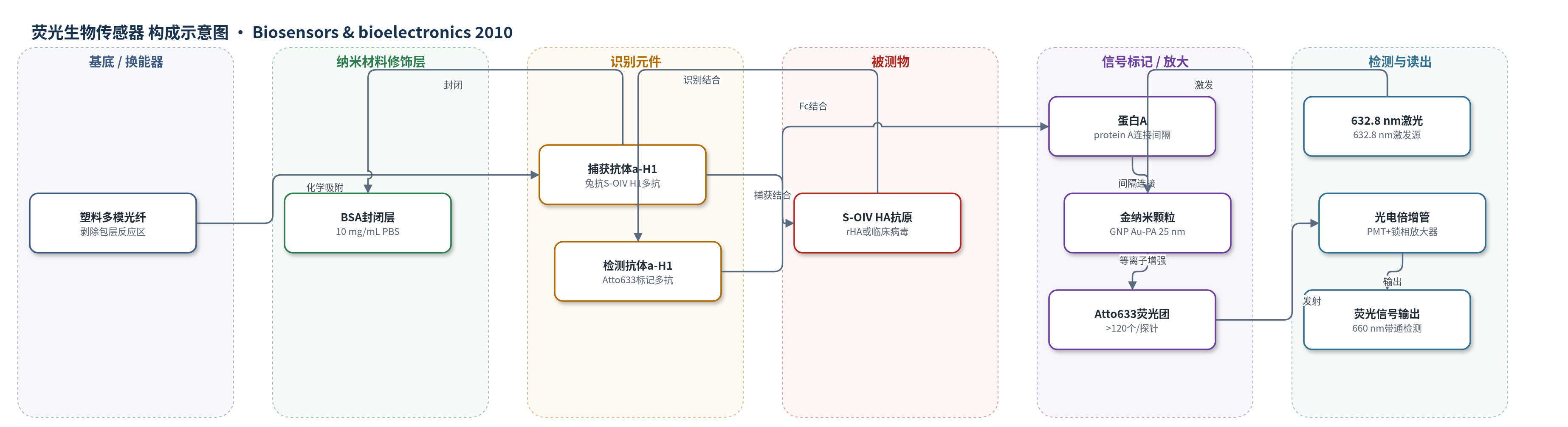
点击图片查看大图 · 依据论文自动绘制
传感器类型
荧光生物传感器
检测对象
猪源流感A(H1N1)病毒(swine-origin influenza A (H1N1) virus, S-OIV)、重组S-OIV H1/血凝素蛋白(recombinant S-OIV H1/HA protein, rHA);样品基质:PBS、健康供体鼻黏膜模拟液(mimic solution)、临床S-OIV培养上清/分离株。
检测原理
光纤剥除区化学吸附兔抗S-OIV H1多克隆抗体(a-H1)作为捕获抗体,并用BSA封闭非特异位点。样品中的S-OIV HA/H1抗原与捕获抗体结合,形成夹心免疫复合物。随后加入LSPCF探针:Atto633荧光标记的抗S-OIV H1检测抗体通过蛋白A连接并吸附在25 nm胶体金纳米颗粒(GNP)表面,再结合到复合物上。632.8 nm激光激发GNP,产生局部表面等离子体(LSP)增强局域电磁场,使GNP附近的Atto633荧光显著增强;每个探针携带>120个Atto633,蛋白A作为间隔避免金属猝灭。增强荧光经660 nm带通滤光片由PMT和锁相放大器检测。S-OIV/HA浓度越高,结合探针越多,荧光信号越强。
检测灵敏度
LOD: 13.9 pg/mL(重组S-OIV H1/rHA);线性范围: 5–50 ng/mL;R^2 = 0.9375;分析灵敏度: 3.7 pg/mL;临床S-OIV LOD: 8.25 × 10^4 copies/mL(PBS);模拟液可检测浓度: 1.65 × 10^5 copies/mL。
效应效果
该传感器对S-OIV具有良好选择性:10倍稀释H5N1病毒培养上清信号与空白对照相同;在健康供体鼻黏膜模拟液中,500倍稀释S-OIV(1.65×10^5 copies/mL)可检出,而10倍稀释H5N1为阴性。与常规捕获ELISA相比,rHA检测LOD由12.2 ng/mL降至13.9 pg/mL,提高10^3倍;临床S-OIV检测LOD由2.06×10^6 copies/mL降至8.25×10^4 copies/mL,提高25倍。与Flu A/B快速试纸条相比,PBS中灵敏度提高约50倍,模拟液中提高约25倍。检测时间约2 h,虽低于实时RT-PCR(10^2–10^3 copies/mL,约6 h),但可数小时内完成抗原诊断,作者认为其高灵敏、高特异,适合临床S-OIV快速诊断并拓展至其他病原微生物检测。
传感器的构成
- 基底/换能器:塑料多模光纤(plastic multimode optical fiber),直径1 mm,剥除包层区作为反应窗口,传导激发光并侧向收集荧光。
- 识别元件:兔抗S-OIV H1多克隆抗体(a-H1 capture antibody),1 μg/mL化学吸附于光纤剥除区,捕获S-OIV HA/H1抗原。
- 封闭层:牛血清白蛋白(BSA, 10 mg/mL PBS),封闭非特异结合位点。
- 检测识别元件:Atto633荧光标记的抗S-OIV H1多克隆抗体(a-H1 detection antibody),识别已捕获的HA抗原。
- 信号标记/纳米材料:胶体金纳米颗粒(GNP, Au–PA, 25 nm)表面吸附蛋白A(protein A)并连接荧光标记检测抗体,形成LSPCF探针,提供局部表面等离子体荧光增强。
- 信号放大元件:Atto633荧光团,每个LSPCF探针携带>120个,在GNP局域电磁场中同步激发增强。
- 读出系统:632.8 nm激光、20×物镜、660 nm带通滤光片、光电倍增管(PMT)和锁相放大器,检测增强荧光。
中文摘要
猪源流感A(H1N1)病毒(S-OIV)于2009年4月被确认为一种新的流感A病毒重配型毒株,并引发流感大流行。准确、及时的诊断对控制流感疫情至关重要。本研究开发了一种局部表面等离子体耦合荧光光纤生物传感器(LSPCF-FOB),将夹心免疫分析技术与局部表面等离子体(LSP)技术相结合,采用针对S-OIV血凝素(HA)蛋白的抗体进行检测。对重组S-OIV H1蛋白的检测限估计为13.9 pg/mL,较使用相同捕获抗体的常规捕获ELISA提高10^3倍。对临床S-OIV分离株的检测,LSPCF-FOB平台计算检测限为8.25×10^4 copies/mL,而常规捕获ELISA为2.06×10^6 copies/mL。此外,与流感A/B快速检测相比,LSPCF-FOB在PBS溶液中对S-OIV的检测限降低约50倍,在以健康供体鼻黏膜为稀释液的模拟液中降低约25倍。研究结果表明,LSPCF-FOB具有高检测灵敏度和特异性,可作为临床S-OIV感染的潜在有效诊断工具,并有望应用于其他临床微生物检测平台的开发。
英文摘要
Swine-origin influenza A (H1N1) virus (S-OIV) was identified as a new reassortant strain of influenza A virus in April 2009 and led to an influenza pandemic. Accurate and timely diagnoses are crucial for the control of influenza disease. We developed a localized surface plasmon coupled fluorescence fiber-optic biosensor (LSPCF-FOB) which combines a sandwich immunoassay with the LSP technique using antibodies against the hemagglutinin (HA) proteins of S-OIVs. The detection limit of the LSPCF-FOB for recombinant S-OIV H1 protein detection was estimated at 13.9 pg/mL, which is 10(3)-fold better than that of conventional capture ELISA when using the same capture antibodies. For clinical S-OIV isolates measurement, meanwhile, the detection limit of the LSPCF-FOB platform was calculated to be 8.25 × 10(4)copies/mL, compared with 2.06 × 10(6)copies/mL using conventional capture ELISA. Furthermore, in comparison with the influenza A/B rapid test, the detection limit of the LSPCF-FOB for S-OIV was almost 50-fold in PBS solution and 25-fold lower in mimic solution, which used nasal mucosa from healthy donors as the diluent. The findings of this study therefore indicate that the high detection sensitivity and specificity of the LSPCF-FOB make it a potentially effective diagnostic tool for clinical S-OIV infection and this technique has the potential to be applied to the development of other clinical microbe detection platforms.