组成图示
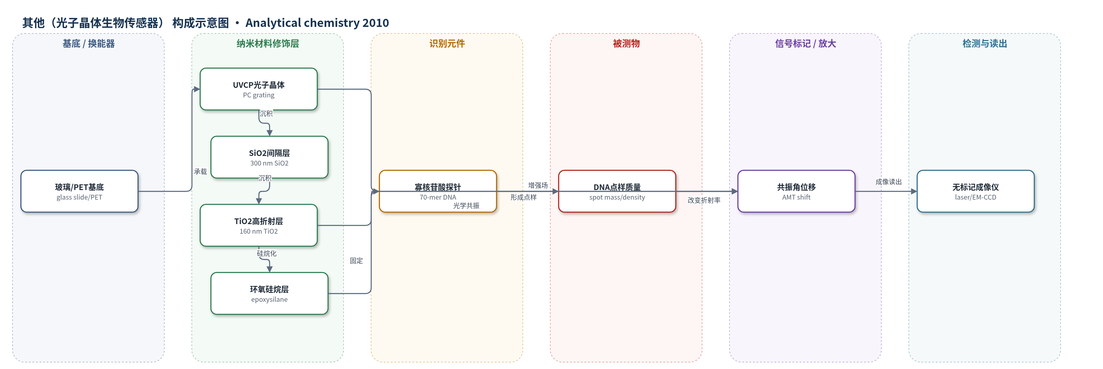
点击图片查看大图 · 依据论文自动绘制
传感器类型
其他(光子晶体生物传感器)
检测对象
DNA 捕获探针点样质量(DNA capture probe spot mass,70-mer oligonucleotide microarray spots);样品基质为点样于光子晶体表面的寡核苷酸微阵列,杂交验证使用大豆子叶总 RNA(soybean cotyledon total RNA,Cy5 标记)
检测原理
该传感器利用一维光子晶体(PC)亚波长光栅的窄带光学共振进行无标记检测。器件由 UVCP 光栅、SiO2 间隔层和 TiO2 高折射率层构成,在固定波长 632.8 nm 下扫描入射角可获得透射最小角(AMT)。点样到环氧硅烷表面的 70-mer 寡核苷酸捕获探针作为生物分子吸附层,其质量密度高于周围空气,使表面附近有效折射率升高,共振角向更大角度位移。由于共振光不能沿表面横向传播,各像素 AMT 相互独立,因此可逐像素拟合透射-角度曲线,生成表面 DNA 质量密度空间图。斑点强度、尺寸、饱和度和局部背景用于质量评分;无酶促或核酸扩增,信号直接随 DNA 点样量变化。
检测灵敏度
缺失点识别灵敏度: 95.2%;特异性: 98.5%;相关系数 r: 未过滤 0.57–0.96,过滤后 0.65–0.96
效应效果
6 张 PC 芯片(两个批次)、32,640 个斑点(含 1300 空白)中,缺失点识别灵敏度 95.2%、特异性 98.5%,96.5% 有意空白被识别。qlf-com 过滤使重复阵列杂交后荧光成对相关系数由 0.57–0.96 升至 0.65–0.96;阈值 0.65 去除 14–28% 斑点,0.7 去除 28–66%,均 p<0.001。器件 σwithin-PC=0.12°。方法无需标记或改流程,适用于 cDNA、寡核苷酸和蛋白微阵列质控。
传感器的构成
- 基底/换能器:玻璃载玻片(glass microscope slide)与柔性聚对苯二甲酸乙二醇酯(PET)基底,承载光子晶体并用于光学成像
- 光子晶体纳米结构:紫外固化聚合物(UVCP)复制的一维周期性亚波长光栅(PC grating),形成窄带光学共振
- 间隔层:300 nm 二氧化硅(SiO2)溅射层,匹配折射率并隔离自荧光 UVCP
- 高折射率层:160 nm 二氧化钛(TiO2)溅射层,增强表面光学共振
- 表面化学修饰层:3-缩水甘油氧基丙基三甲氧基硅烷(epoxysilane, 3-glycidoxypropyltrimethoxysilane),经 O2 等离子体清洁后气相硅烷化,提供环氧基团固定 DNA
- 识别元件:点样固定的 70-mer 寡核苷酸捕获探针(DNA oligonucleotide capture probes,大豆基因),作为微阵列识别元件和被测 DNA 质量
- 信号标记物:无标记成像不依赖荧光标记;杂交验证使用 Cy5-dUTP 标记的大豆总 RNA(Cy5-labeled target RNA)
- 封闭剂:杂交前使用牛血清白蛋白(BSA)封闭
中文摘要
DNA 微阵列捕获探针点样过程中的技术变异是获得高质量微阵列数据的重要障碍。现有荧光标记方法虽可在杂交前可视化点样阵列,但通常需要改变标准微阵列流程或引入标记/脱色步骤。本研究采用光子晶体(PC)生物传感器表面和高分辨率无标记成像检测仪器,在不改变标准点样与杂交流程的前提下,对点样寡核苷酸微阵列进行杂交前成像。通过监测 PC 光学共振条件随表面生物分子吸附的变化,获得斑点强度、尺寸、饱和度和局部背景强度,并据此自动识别因机械故障或样品耗尽导致的缺失点,同时为每个点样特征分配反映质量的综合评分。缺失点识别灵敏度超过 95%。基于斑点质量评分进行过滤后,重复阵列杂交后斑点荧光强度的成对相关系数提高,表明无标记斑点质量评分能够捕获微阵列数据中的变异。该成像模式可用于印刷 cDNA、寡核苷酸和蛋白微阵列的质量控制。
英文摘要
Technical variability during DNA capture probe printing remains an important obstacle to obtaining high quality data from microarray experiments. While methods that use fluorescent labels for visualizing printed arrays prior to hybridization have been presented, the ability to measure spot density using label-free techniques would provide valuable information on spot quality without altering standard microarray protocols. In this study, we present the use of a photonic crystal biosensor surface and a high resolution label-free imaging detection instrument to generate prehybridization images of spotted oligonucleotide microarrays. Spot intensity, size, level of saturation, and local background intensity were measured from these images. This information was used for the automated identification of missed spots (due to mechanical failure or sample depletion) as well as the assignment of a score that reflected the quality of each printed feature. Missed spots were identified with >95% sensitivity. Furthermore, filtering based on spot quality scores increased pairwise correlation of posthybridization spot intensity between replicate arrays, demonstrating that label-free spot quality scores captured the variability in the microarray data. This imaging modality can be applied for the quality control of printed cDNA, oligonucleotide, and protein microarrays.