组成图示
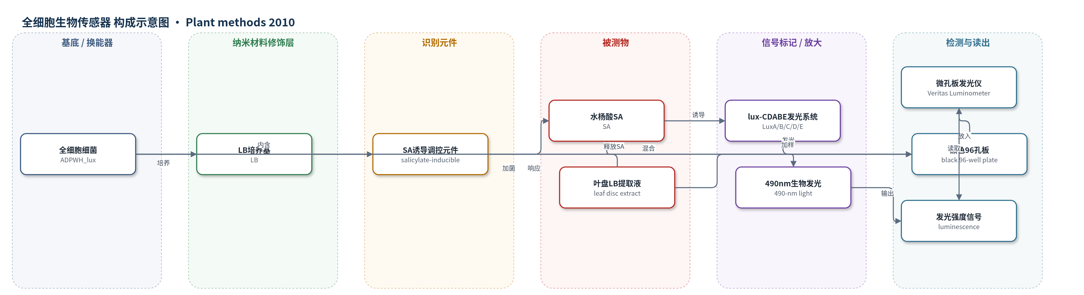
点击图片查看大图 · 依据论文自动绘制
传感器类型
全细胞生物传感器
检测对象
水杨酸(salicylic acid, SA);样品基质:病原感染拟南芥叶片叶盘 LB 提取液
检测原理
病原感染拟南芥叶盘在 LB 中 95°C 煮沸 20 min,使细胞内水杨酸(SA)释放到提取液。提取液与 Acinetobacter sp. ADPWH_lux 全细胞生物传感器混合后,SA 进入细菌并激活染色体整合的 SA 诱导型 lux-CDABE 操纵子。该操纵子表达荧光素酶 LuxA/LuxB 以及底物合成酶 LuxC/LuxD/LuxE,使细胞内合成发光底物并催化其氧化,产生 490 nm 生物发光。SA 浓度越高,lux 基因簇诱导越强,发光强度越大。孵育 1 h 后用微孔板发光仪读取发光信号,从而以生物发光强度估计样品中 SA 水平。
检测灵敏度
原文未报告LOD、线性范围、灵敏度斜率或相关系数。
效应效果
该传感器对 SA、methyl-SA 和 acetylsalicylic acid 高度特异,适合粗植物提取液检测。直接煮沸法可区分病原感染 Col-0、npr1 和 sid2 的 SA 差异,结果与原有生物传感器法和 HPLC 一致,并省去称重、研磨和离心。高通量格式采用 96 孔托盘、96 孔 PCR 板和 96 孔培养板,已筛选约 10000 株 M2 植株,初筛 35 个候选 npr1 抑制子,复筛确认 6 个感染后 SA 低于 npr1 的突变体。HPLC 验证 62 和 69 的游离及总 SA 极低,类似 eds5 和 sid2,并证明二者为 eds5 等位。病原接种显示 34、46、49、79 中 Psm ES4326 生长显著高于 npr1-3,其中 49 高 10 倍。作者认为该方法成本低、通量高,可用于 SA 代谢突变体筛选。
传感器的构成
- 全细胞换能器:Acinetobacter sp. ADPWH_lux 细菌细胞,作为 SA 识别与生物发光信号转导单元
- 识别元件:染色体整合的 SA 诱导型 lux-CDABE 操纵子调控元件,响应 SA、methyl-SA 和 acetylsalicylic acid
- 信号标记物:lux-CDABE 基因簇,编码荧光素酶 LuxA/LuxB 及底物合成酶 LuxC/LuxD/LuxE,产生 490 nm 生物发光
- 反应介质:LB 培养基,用于生物传感器培养及叶盘 SA 释放与稀释
- 信号读出:Veritas Microplate Luminometer,检测 490 nm 生物发光强度
中文摘要
水杨酸(SA)是植物抵御生物营养型病原菌的关键防御信号分子,准确测定植物体内 SA 水平对解析 SA 介导的免疫反应至关重要。虽然 HPLC 和 GC/MS 常用于 SA 定量,但成本高且耗时。作者此前报道了基于 Acinetobacter sp. ADPWH_lux 细菌生物传感器的快速 SA 定量方法,可实现高通量分析。本研究进一步简化样品制备流程,提出一种高通量分离 SA 代谢突变体的策略。该方法将病原感染叶片叶盘直接置于 LB 培养基中煮沸,使 SA 释放到提取液中,再与生物传感器共孵育并测定生物发光,从而避免称重、研磨和离心等耗时步骤。直接煮沸法能够区分病原感染野生型、npr1 和 sid2 植株间的 SA 水平差异,结果与原有生物传感器法和 HPLC 法一致。作者将该流程适配为 96 孔高通量格式,筛选约 10000 株 M2 突变体,鉴定出 6 个感染后 SA 积累低于 npr1 的抑制子,其中 2 个与 eds5 等位,另 4 个对 Pseudomonas syringae pv. maculicola ES4326 更敏感。该快速 SA 估计方法可显著降低成本和时间,适用于 SA 代谢突变体遗传筛选及相关酶功能表征。
英文摘要
BACKGROUND: Salicylic acid (SA) is a key defense signal molecule against biotrophic pathogens in plants. Quantification of SA levels in plants is critical for dissecting the SA-mediated immune response. Although HPLC and GC/MS are routinely used to determine SA concentrations, they are expensive and time-consuming. We recently described a rapid method for a bacterial biosensor Acinetobacter sp. ADPWH_lux-based SA quantification, which enables high-throughput analysis. In this study we describe an improved method for fast sample preparation, and present a high-throughput strategy for isolation of SA metabolic mutants.
RESULTS: On the basis of the previously described biosensor-based method, we simplified the tissue collection and the SA extraction procedure. Leaf discs were collected and boiled in Luria-Bertani (LB), and then the released SA was measured with the biosensor. The time-consuming steps of weighing samples, grinding tissues and centrifugation were avoided. The direct boiling protocol detected similar differences in SA levels among pathogen-infected wild-type, npr1 (nonexpressor of pathogenesis-related genes), and sid2 (SA induction-deficient) plants as did the previously described biosensor-based method and an HPLC-based approach, demonstrating the efficacy of the protocol presented here. We adapted this protocol to a high-throughput format and identified six npr1 suppressors that accumulated lower levels of SA than npr1 upon pathogen infection. Two of the suppressors were found to be allelic to the previously identified eds5 mutant. The other four are more susceptible than npr1 to the bacterial pathogen Pseudomonas syringae pv. maculicola ES4326 and their identity merits further investigation.
CONCLUSIONS: The rapid SA extraction method by direct boiling of leaf discs further reduced the cost and time required for the biosensor Acinetobacter sp. ADPWH_lux-based SA estimation, and allowed the screening for npr1 suppressors that accumulated less SA than npr1 after pathogen infection in a high-throughput manner. The highly efficacious SA estimation protocol can be applied in genetic screen for SA metabolic mutants and characterization of enzymes involved in SA metabolism. The mutants isolated in this study may help identify new components in the SA-related signaling pathways.