组成图示
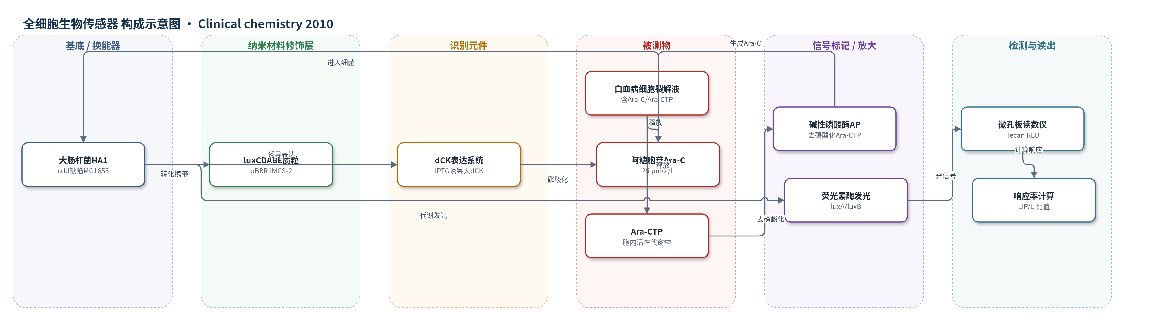
点击图片查看大图 · 依据论文自动绘制
传感器类型
全细胞生物传感器
检测对象
阿糖胞苷(cytarabine, Ara-C)、阿糖胞苷三磷酸(Ara-CTP);样品基质:AML患者骨髓/外周血单核细胞裂解液、AML细胞系培养裂解液
检测原理
白血病细胞与Ara-C孵育后,Ara-C经hENT1进入细胞,由dCK磷酸化为Ara-CMP,并进一步磷酸化为活性形式Ara-CTP。细胞裂解后,裂解液同时释放Ara-C和Ara-CTP。加入IPTG诱导表达人dCK的发光大肠杆菌HA1后,Ara-C可进入细菌并被磷酸化,干扰细菌DNA合成与代谢,使luxCDABE荧光素酶催化FMNH2和长链醛氧化,发射蓝绿光。Ara-CTP不能进入细菌;加入AP后,Ara-CTP被去磷酸化为Ara-C,从而产生额外发光。通过比较加AP与不加AP的峰值发光(LIP/LI),可定量反映裂解液中Ara-CTP浓度,即白血病细胞将Ara-C转化为活性代谢物的能力,进而预测药物敏感性。
检测灵敏度
LOD: 0.025 μmol/L(25 nmol/L)磷酸化 Ara-C/Ara-CTP,显著增加光输出(P < 0.05)
效应效果
该传感器对Ara-C具有选择性,0.1 μmol/L Ara-C即可引起显著发光增加,而对嘌呤类似物fludarabine无响应。Ara-CTP在12.5–100 nmol/L及0–100 μmol/L范围内与加/不加AP的发光响应相关,25 nmol/L可被显著检测。KG-1a细胞对Ara-C敏感,THP-1部分耐药,8小时检测与3天Cell Titer-Glo细胞毒性试验密切相关。回顾性24份AML骨髓或外周血样本中,12例反应患者生物传感器响应为21–128%(中位55%),12例无反应患者为-7–6%(中位0%),可在8小时内预测临床反应。作者认为该检测有望用于化疗前快速评估Ara-C敏感性,降低敏感患者剂量,并开发为临床诊断试剂盒。
传感器的构成
- 细胞基底/换能器:大肠杆菌MG1655衍生cdd缺陷突变体E. coli HA1,作为全细胞传感与发光平台,cdd缺陷避免Ara-C去氨失活,pyrE缺陷增强Ara-C毒性响应
- 识别/代谢元件:IPTG诱导表达的人脱氧胞苷激酶dCK(pTrcHUMdCK/pTrc99-A质粒),将Ara-C磷酸化为Ara-CMP并进一步生成Ara-CTP,介导药物毒性识别
- 信号报告元件:pBBR1MCS-2质粒携带Photorhabdus luminescens luxCDABE操纵子,编码luxA/luxB荧光素酶及luxC/luxD/luxE脂肪酸还原/酰基转移/酰基蛋白合成酶,产生自发光
- 诱导剂:IPTG,诱导dCK表达,使细菌对Ara-C产生发光响应
- 样品裂解体系:EDTA 1.5 mmol/L与皂苷saponin 1.0 g/L,裂解白血病细胞并释放胞内Ara-C和Ara-CTP
- 信号区分酶:碱性磷酸酶AP 10 U,将Ara-CTP去磷酸化为Ara-C,用于区分游离Ara-C与胞内Ara-CTP
- 读出系统:Tecan多模式微孔板读数仪,监测生物发光RLU和600 nm吸光度生长
中文摘要
阿糖胞苷(Ara-C)是治疗急性髓系白血病(AML)的关键药物,但最多30%患者治疗无反应,因此需要治疗前筛选患者血样以预测药物反应。本研究通过转座突变构建胞苷脱氨酶(cdd)缺陷的大肠杆菌MG1655突变体,使其对Ara-C敏感,并转化携带Photorhabdus luminescens luxCDABE操纵子的质粒,构建自发光全细胞生物传感器E. coli HA1。该菌株在IPTG诱导下表达人脱氧胞苷激酶(dCK),用于8小时检测白血病细胞对Ara-C的摄取和磷酸化。结果显示,胞内0.025 μmol/L磷酸化Ara-C可被显著增加的光输出检测(P<0.05)。已知对Ara-C敏感或耐药的AML细胞系中,8小时检测与3天细胞毒性试验密切相关。回顾性检测24份骨髓或外周血临床样本,该生物传感器检测可在8小时内预测白血病细胞对Ara-C的反应。结论认为,该检测可作为化疗前评估AML细胞Ara-C敏感性的预测工具,有助于对敏感患者降低Ara-C剂量;其监测胞内Ara-CTP水平,若充分验证可用于临床。
英文摘要
BACKGROUND: The nucleoside analog cytarabine (Ara-C [cytosine arabinoside]) is the key agent for treating acute myeloid leukemia (AML); however, up to 30% of patients fail to respond to treatment. Screening of patient blood samples to determine drug response before commencement of treatment is needed. This project aimed to construct and evaluate a self-bioluminescent reporter strain of Escherichia coli for use as an Ara-C biosensor and to design an in vitro assay to predict Ara-C response in clinical samples.
METHODS: We used transposition mutagenesis to create a cytidine deaminase (cdd)-deficient mutant of E. coli MG1655 that responded to Ara-C. The strain was transformed with the luxCDABE operon and used as a whole-cell biosensor for development an 8-h assay to determine Ara-C uptake and phosphorylation by leukemic cells.
RESULTS: Intracellular concentrations of 0.025 μmol/L phosphorylated Ara-C were detected by significantly increased light output (P < 0.05) from the bacterial biosensor. Results using AML cell lines with known response to Ara-C showed close correlation between the 8-h assay and a 3-day cytotoxicity test for Ara-C cell killing. In retrospective tests with 24 clinical samples of bone marrow or peripheral blood, the biosensor-based assay predicted leukemic cell response to Ara-C within 8 h.
CONCLUSIONS: The biosensor-based assay may offer a predictor for evaluating the sensitivity of leukemic cells to Ara-C before patients undergo chemotherapy and allow customized treatment of drug-sensitive patients with reduced Ara-C dose levels. The 8-h assay monitors intracellular Ara-CTP (cytosine arabinoside triphosphate) levels and, if fully validated, may be suitable for use in clinical settings.