组成图示
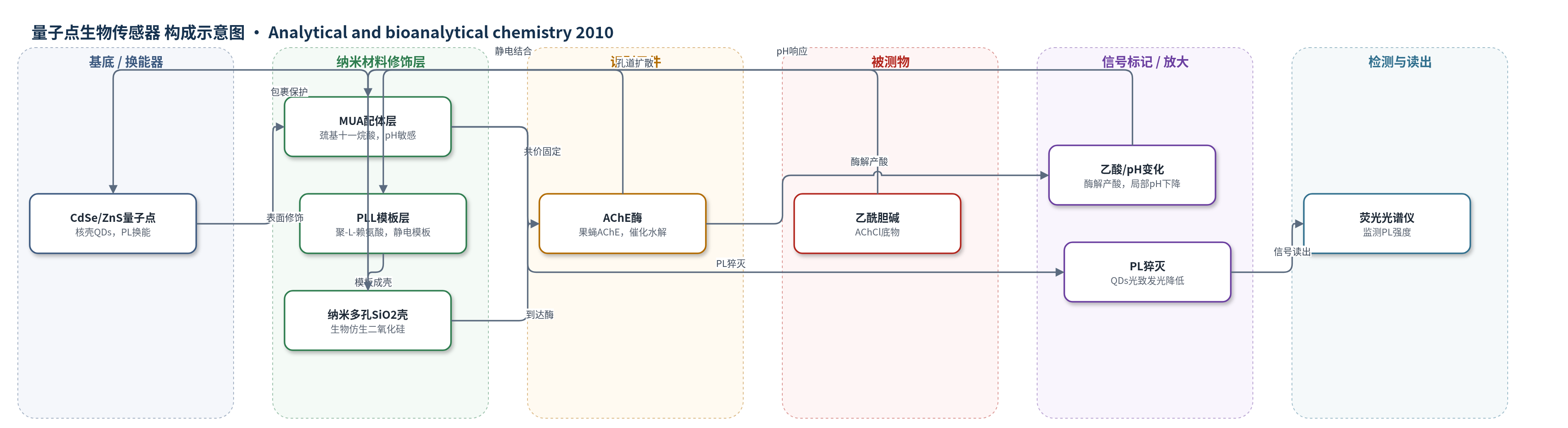
点击图片查看大图 · 依据论文自动绘制
传感器类型
量子点生物传感器
检测对象
乙酰胆碱氯化物(acetylcholine chloride,AChCl),缓冲水溶液;乙酰胆碱酯酶抑制剂(AChE inhibitors,潜在应用)
检测原理
该传感器以AChE作为识别与催化元件,以MUA包覆的CdSe/ZnS量子点作为pH敏感光致发光换能器。乙酰胆碱氯化物通过生物仿生二氧化硅壳的纳米孔扩散至内部,与AChE活性中心接触并被水解为胆碱和乙酸。酶催化反应在QDs周围产生局部酸化,使MUA配体羧基的质子化状态和QDs表面界面环境改变,从而引起量子点光致发光猝灭。底物浓度越高,单位时间内生成的乙酸越多,局部pH下降越明显,PL信号降低越大。该过程利用酶催化反复转化底物实现化学放大,无需额外核酸或纳米酶放大策略,最终通过荧光光谱仪读取PL强度变化。
检测灵敏度
LOD: 1 μM;线性范围: 100–1,000 μM
效应效果
论文未报告选择性、抗干扰、重现性RSD、实际样品加标回收率或与ELISA/HPLC/qPCR等方法的对比。稳定性方面,QD/AChE/PLL/silica生物传感器在5天后仍保持100%剩余活性,45天后保持65%;未包埋于生物仿生二氧化硅的QD/AChE复合物在20天后活性仅降至50%。作者指出储存寿命超过2个月。生物仿生二氧化硅壳可保护AChE免受展开、变性和蛋白酶攻击,同时降低CdSe/ZnS量子点有毒Cd2+的泄漏风险,并允许底物自由扩散。作者主张该平台可用于溶液中低浓度底物监测,并为检测乙酰胆碱酯酶抑制剂提供稳定、灵敏的纳米生物传感器基础。
传感器的构成
- 换能器/信号标记:CdSe/ZnS核壳量子点(QDs),提供光致发光信号并作为pH敏感光学换能器
- 表面配体/偶联层:巯基十一烷酸(MUA),提供羧基用于EDC/NHS共价偶联AChE,并赋予QDs pH敏感光致发光
- 识别/催化元件:果蝇乙酰胆碱酯酶(Dm. AChE),催化乙酰胆碱水解并产生乙酸
- 模板/稳定层:聚-L-赖氨酸(PLL),正电荷模板,静电结合AChE并引导生物仿生二氧化硅形成
- 外壳/稳定层:生物仿生纳米多孔二氧化硅(silica,由TMOS水解硅酸形成),包裹QD/AChE/PLL,允许底物扩散、阻挡蛋白酶并稳定QDs与酶
中文摘要
CdSe/ZnS核壳量子点(QDs)经巯基十一烷酸(MUA)功能化后,以聚-L-赖氨酸(PLL)为模板在其外表面形成生物仿生二氧化硅壳,构成用于光学生物传感器的换能与稳定体系。果蝇乙酰胆碱酯酶(AChE)在二氧化硅形成过程中被共价固定于QDs表面,作为相对不稳定酶的原理验证模型。所得QD/AChE/PLL/silica纳米生物传感器中,PLL模板二氧化硅外壳不改变QDs的光学性质,并能保护酶免受展开和变性。二氧化硅壳的小孔允许分析物自由扩散至酶活性中心,同时阻止蛋白酶接近酶。通过监测QDs光致发光随酶反应引起局部pH变化的响应,评估该纳米生物传感器对底物乙酰胆碱氯化物的检测性能;pH变化与溶液中分析物浓度相关。该生物检测体系稳定,储存寿命超过2个月,为利用该纳米结构生物传感器检测乙酰胆碱酯酶抑制剂提供了基础。
英文摘要
CdSe/ZnS core/shell quantum dots (QDs) are functionalized with mercaptoundecanoic acid (MUA) and subsequently covered with poly-L-lysine (PLL) as the template for the formation of the silica outer shell. This nanocomposite is used as a transduction and stabilization system for optical biosensor development. The covalent immobilization of the enzyme acetylcholinesterase from Drosophila melanogaster (AChE) during the formation of the biomimetically synthesized silica is used here as a model, relatively unstable enzyme, as a proof of principle. The enzyme is successfully immobilized onto the QDs and then stabilized by the PLL capping and the subsequent formation of the outer nanoporous silica thin shell, giving rise to the QD/AChE/PLL/silica biosensor. It is shown that the poly-L-lysine templated silica outer shell does not modify the optical properties of the quantum dots, while it protects the enzyme from unfolding and denaturation. The small pores of the silica shell allow for the free diffusion of the analyte to the active center of the enzyme, while it does not allow for the proteases to reach the enzyme. The response of the QD/AChE/PLL/silica nano-biosensor to its substrate, acetylcholine chloride, is evaluated by monitoring the changes in the QDs' photoluminescence which are related to the changes in pH. These pH changes of the surrounding environment of the QDs are induced by the enzymatic reaction, and are associated with the analyte concentration in the solution. The biodetection system proposed is shown to be stable with a storage lifetime of more than 2 months. The data presented provides the grounds for the application of this nanostructured biosensor for the detection of AChE inhibitors.