组成图示
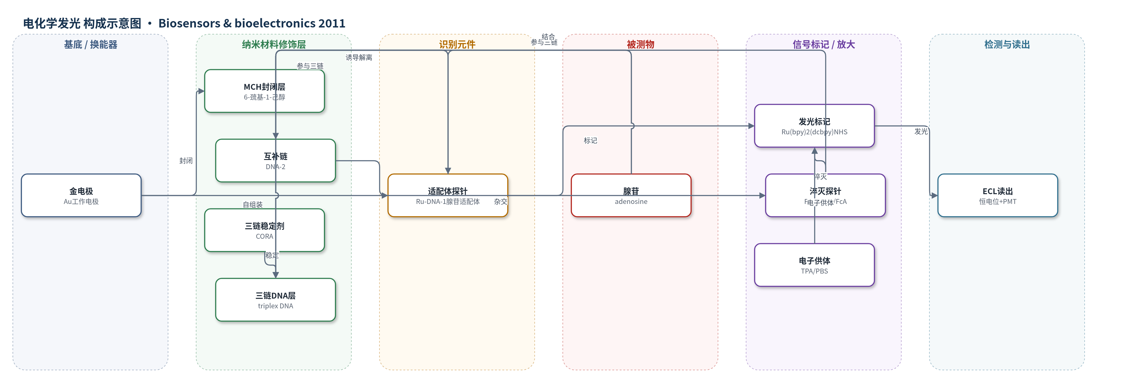
点击图片查看大图 · 依据论文自动绘制
传感器类型
电化学发光(ECL)生物传感器
检测对象
腺苷(adenosine);样品基质:PBS缓冲液、20%人血清
检测原理
金电极表面自组装Ru-DNA-1腺苷适配体探针,MCH封闭后,DNA-2与Fc-DNA-3在CORA存在下形成三链DNA。Fc-DNA-3上的FcA通过电子转移淬灭Ru(bpy)3^2+的电化学发光,使背景信号较低。加入腺苷后,适配体优先与腺苷结合并形成发夹构象的适配体-腺苷复合物,引发Ru-DNA-1结构开关,三链结构解离,Fc-DNA-3脱离电极表面,淬灭作用减弱。在0.10 mol L−1 PBS和0.10 mol L−1 TPA中,于+1.00 V恒电位下,Ru(bpy)3^2+被氧化激发并与TPA反应产生ECL,发光强度增量ΔI随腺苷浓度升高而增大。三链结构更紧密,有利于电荷转移淬灭,从而提高信噪比和特异性。
检测灵敏度
LOD: 2.7 × 10⁻¹⁰ mol L−1;线性范围: 1.0 × 10⁻⁹–1.0 × 10⁻⁸ mol L−1;回归方程: Y = 17.167 + 91.253X(X单位10−9 mol L−1,Y为ΔIECL);R = 0.999。对照ECL-duplex: LOD: 2.3 × 10⁻⁹ mol L−1;线性范围: 1.0 × 10⁻⁸–1.0 × 10⁻⁷ mol L−1。
效应效果
该传感器对腺苷选择性良好,10−8 mol L−1胞苷和尿苷未引起显著ECL变化。CORA用于提高三链结构稳定性。与ECL-duplex对照相比,三链传感器空白值更低,灵敏度约提高1个数量级,并显著提升特异性。在20%人血清中检测腺苷,回收率为90%–100%,变异系数小于10%。作者指出其检出限比无标记无试剂适配体传感器低10000倍,比金纳米颗粒聚集比色法低6个数量级以上,比二茂铁标记适配体探针低100倍,比SPR传感器低1个数量级,显示用于腺苷检测的应用潜力。
传感器的构成
- 基底/换能器电极:金电极(Au electrode,直径1.5 mm),作为工作电极和ECL换能器
- 识别探针自组装层:巯基化腺苷适配体DNA-1自组装于金表面,5′端标记Ru(bpy)2(dcbpy)NHS,形成ECL发光探针Ru-DNA-1
- 封闭层:6-巯基-1-己醇(MCH)封闭未覆盖金表面,减少非特异性吸附
- 互补链杂交层:DNA-2与Ru-DNA-1互补杂交,形成双链结构
- 三链DNA结构层:DNA-2与Fc-DNA-3杂交,并在珊瑚碱盐酸盐(CORA)存在下与适配体三链特征链形成三链DNA
- 淬灭探针层:Fc-DNA-3 5′端标记二茂铁单羧酸(FcA),作为ECL淬灭剂
- ECL反应介质/电子供体:0.10 mol L−1 PBS(pH 7.4)含0.10 mol L−1三丙胺(TPA),在+1.00 V下激发Ru(bpy)3^2+发光
中文摘要
本文设计了一种基于三链DNA的电化学发光(ECL)生物传感器,用于检测腺苷。该传感器以腺苷适配体作为分子识别元件,利用二茂铁单羧酸(FcA)淬灭三(2,2′-联吡啶)钌(Ru(bpy)3^2+)的ECL信号。通过自组装技术,将巯基化发夹腺苷适配体探针标记ECL发光体Ru(bpy)3^2+衍生物(Ru-DNA-1)固定于金电极表面。在珊瑚碱盐酸盐(CORA)存在下,适配体中的三链特征链与DNA-2和Fc-DNA-3形成三链DNA结构,其中Fc-DNA-3携带ECL淬灭剂FcA。加入腺苷后,适配体序列优先与腺苷形成发夹构象的适配体-腺苷复合物,引发DNA-1结构开关,并因淬灭探针解离而产生强ECL信号。同时构建了ECL双链DNA传感器作为对照。ECL三链DNA传感器和ECL双链DNA传感器的检出限分别为2.7×10⁻¹⁰ mol L−1和2.3×10⁻⁹ mol L−1,表明三链传感器显著提高了灵敏度和特异性。
英文摘要
A novel electrogenerated chemiluminescence (ECL) biosensor based on the construction of triplex DNA for the detection of adenosine was designed. The ECL biosensor employs an aptamer as a molecular recognition element, and quenches ECL of tris(2,2'-bipyridine) ruthenium (Ru(bpy)(3)(2+)) by ferrocenemonocarboxylic acid (FcA). Through self-assembly technology, the ECL probe of thiolated hairpin adenosine aptamer tagged was self-assembled onto the surface of a gold electrode with an ECL signal producer Ru(bpy)(3)(2+) derivative (Ru-DNA-1). The adenosine aptamer, including a section of triplex characteristic chain, formatted triplex DNA with two other DNAs (DNA-2, Fc-DNA-3) in the presence of triplex DNA binder coralyne chloride (CORA). Fc-DNA-3 was tagged with an ECL quencher ferrocenemonocarboxylic acid (FcA), a quenching probe. In the presence of adenosine, the aptamer sequence (Ru-DNA-1) prefers to form the aptamer-adenosine complex with hairpin configuration and the switch of the DNA-1 occurs in conjunction with the generation of a strong ECL signal owing to the dissociation of a quenching probe. Meanwhile, a control experiment was performed; the ECL-duplex biosensor was designed to detect adenosine. The detection limits were 2.7×10(-10) mol L(-1) and 2.3×10(-9) mol L(-1) for the ECL-triplex DNA biosensor and ECL-duplex DNA biosensor, respectively, which demonstrated that the ECL-triplex DNA biosensor improved the sensitivity and specificity greatly.