组成图示
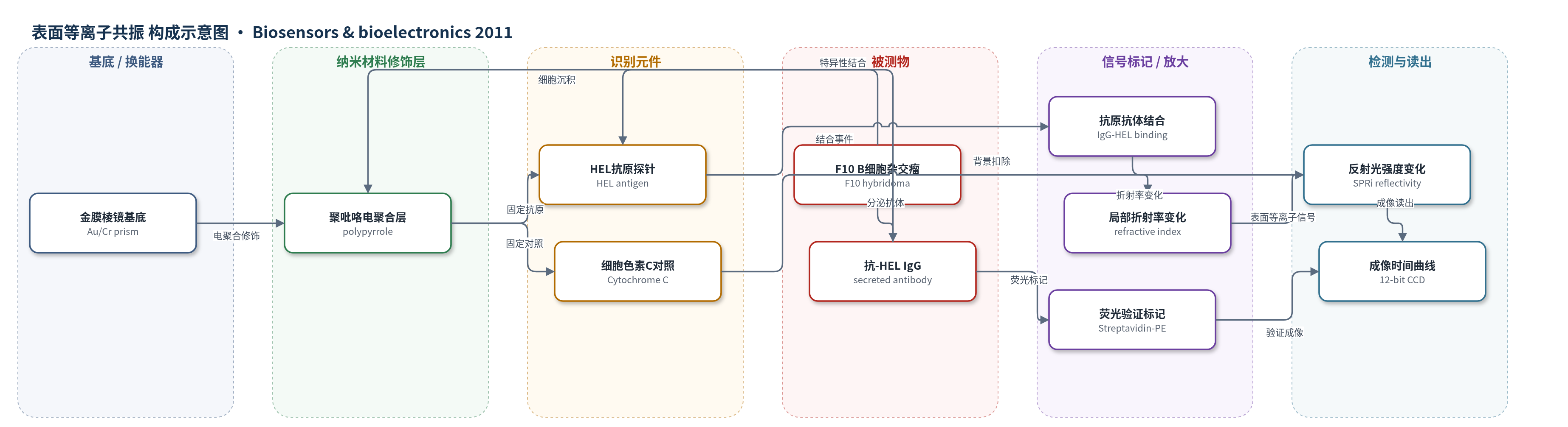
点击图片查看大图 · 依据论文自动绘制
传感器类型
表面等离子共振(SPR)生物传感器
检测对象
抗-HEL 免疫球蛋白(anti-HEL immunoglobulin G, IgG),样品基质:B 细胞杂交瘤培养上清/芯片局部培养基(serum-free RPMI)
检测原理
该传感器以金膜棱镜为 SPR 基底,通过吡咯/蛋白电共聚物化在表面固定 HEL 抗原探针和细胞色素 C 对照。F10 B 细胞杂交瘤加入 PEEK 培养腔后沉积于敏感表面附近,持续分泌抗-HEL IgG。分泌的 IgG 与表面 HEL 结合,使金膜附近数百纳米范围内的折射率改变,SPR 反射光强度随之变化,由 CCD 实时成像。由于细胞贴近表面,局部抗体浓度高于主体培养基,因此可在数分钟内检测到低于纳摩尔体相浓度的分泌事件。信号随结合量增加而上升,探针饱和后出现平台;归一化后动力学与探针密度无关,主要反映分泌过程。
检测灵敏度
原文未报告明确 LOD、线性范围、灵敏度斜率或 R^2。
效应效果
实验显示 HEL 探针点反射率随孵育时间增加,而细胞色素 C 对照无明显漂移,表明识别特异、背景低;SPR 后荧光标记进一步确认表面结合的是抗-HEL IgG。与 ELISA 相比,SPRi 在数分钟内即可出现显著信号,而 ELISA 需 30–60 min 终点分析;ELISA 30 min 上清抗体浓度为 51.3 ± 5.6 ng/mL,8 h 达 579.1 ± 47.7 ng/mL。SPRi 短时呈线性、长时呈指数并达到平台,归一化后不同探针浓度曲线重叠,特征时间与 ELISA 一致且与探针密度无关。该方法无标记、一步法、可多探针并行,适合免疫细胞分泌表型分析。
传感器的构成
- 基底/换能器:50 nm 金膜/2 nm 铬膜棱镜(Au/Cr prism),提供 SPR 传感表面并作为工作电极
- 修饰层:吡咯/蛋白电共聚物化聚吡咯微阵列(polypyrrole microarray),通过 NHS-pyrrole 偶联蛋白与自由吡咯共聚合固定探针
- 识别元件:HEL 抗原探针(Hen Egg Lysozyme, HEL),捕获细胞分泌的抗-HEL IgG
- 对照探针:细胞色素 C(Cytochrome C),用于评估非特异吸附和背景扣除
- 样品/细胞:F10 B 细胞杂交瘤(F10 hybridoma),持续分泌抗-HEL 单克隆 IgG
- 培养介质/表面饱和:RPMI 培养基(含 HEPES)及 10% FBS,用于细胞加载和表面饱和
- 读出系统:SPRi 成像仪与 12-bit CCD,监测反射光强度变化并生成时间曲线
- 验证标记:生物素化抗小鼠抗体与链霉亲和素-藻红蛋白(Streptavidin-Phycoerythrin),用于 SPR 后荧光确认
中文摘要
细胞分泌分子对基础研究和医学应用均具有重要意义。本研究提出一种基于生物芯片的方法,用于瞬时监测体外培养 B 淋巴细胞分泌的蛋白。作者利用表面等离子共振成像(SPRi)对蛋白生物芯片进行成像,芯片上微阵列化固定了抗原蛋白鸡卵白蛋白/卵溶菌酶(HEL)及一系列对照蛋白。将 B 细胞杂交瘤培养于芯片上,可实时监测其分泌的 HEL 特异性免疫球蛋白(抗体),并在数分钟内检测到,而标准 ELISA 通常需要 30–60 min 孵育。该快速、敏感检测得益于细胞沉积在生物芯片敏感表面附近,使局部抗体浓度在稀释到主体培养基前显著升高。此外,SPR 响应经归一化后与表面固定探针密度无关。该生物传感器有望为其他免疫细胞分泌蛋白的定性和半定量分析提供新工具。
英文摘要
The secretions of molecules by cells are of tremendous interest for both fundamental insights studies and medical purposes. In this study, we propose a new biochip-based approach for the instantaneous monitoring of protein secretions, using antibody production by B lymphocytes cultured in vitro. This was possible thanks to the Surface Plasmon Resonance imaging (SPRi) of a protein biochip where antigen proteins (Hen Egg Lysozyme, HEL) were micro-arrayed along with series of control proteins. B cell hybridomas were cultured on the chip and the secretion of immunoglobulins (antibody) specific to HEL was monitored in real-time and detected within only few minutes rather than after a 30-60 min incubation with standard ELISA experiments. This fast and sensitive detection was possible thanks to the sedimentation of the cells on the biochip sensitive surface, where local antibody concentrations are much higher before dilution in the bulk medium. An other interesting feature of this approach for the secretion monitoring was the independence of the SPR response--after normalization--regarding to the density of the surface-immobilized probes. Such biosensor might thus pave the way to new tools capable of both qualitative and semi-quantitative analysis of proteins secreted by other immune cells.