组成图示
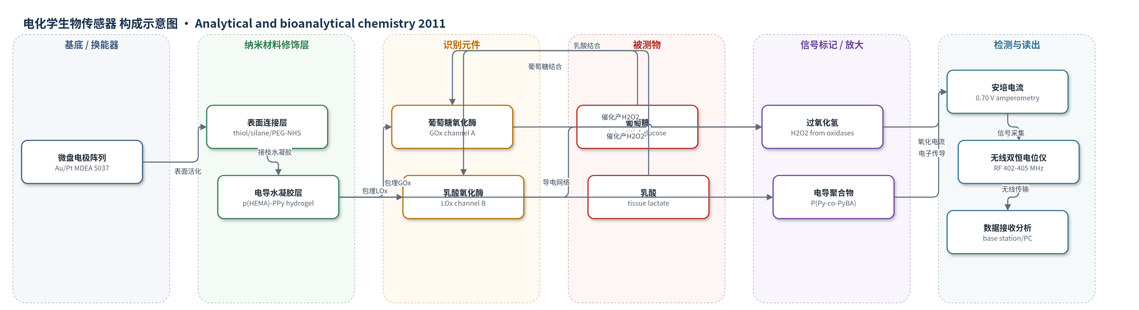
点击图片查看大图 · 依据论文自动绘制
传感器类型
电化学生物传感器
检测对象
葡萄糖(glucose)、乳酸(lactate);样品基质:肌内组织/间质液(intramuscular tissue/interstitial fluid),体外PBS
检测原理
葡萄糖和乳酸从肌内组织/间质液扩散进入电导水凝胶层。葡萄糖通道中,葡萄糖氧化酶(GOx)催化葡萄糖氧化生成葡萄糖酸内酯和过氧化氢;乳酸通道中,乳酸氧化酶(LOx)催化乳酸氧化生成丙酮酸和过氧化氢。生成的H2O2在微盘工作电极上于0.70 V(vs Ag/AgCl)发生氧化,释放电子,形成安培电流。P(Py-co-PyBA)电导聚合物网络提供电子传导路径并抑制干扰物氧化,使电流与底物浓度呈线性关系。双通道分别采集葡萄糖和乳酸信号,经无线双恒电位仪数字化并遥测传输。
检测灵敏度
LOD: 0.2 mM (glucose), 0.4 mM (lactate);线性范围: 0.1–13.0 mM (glucose), 1.0–7.0 mM (lactate);灵敏度: 0.59 μA/mM (glucose), 2.11 μA/mM (lactate)
效应效果
体外细胞实验显示两种水凝胶无细胞毒性,HA-VSMC活力>80%;PPy基电导水凝胶使RMS13细胞密度增加81%、PC12增加12%(p=0.05),并抑制干扰物氧化。含MPC水凝胶降低蛋白吸附,水化率与抗蛋白吸附相关R2=89%;大鼠体内2周植入后,1 mol% MPC样品仅形成薄包膜并减少炎症,无肉芽肿反应。双恒电位仪dummy cell测试通道误差为6.5%(10 MΩ/1 μF)和0.6%(10 MΩ)。37 °C连续运行5天后保持初始响应80%。SD大鼠出血模型中,肌内乳酸较系统乳酸更快升高,与血气/代谢分析仪测得系统乳酸不一致。作者认为该芯片可用于创伤出血复苏、ICU连续监测和战场/灾害场景。
传感器的构成
- 基底/换能器电极:硼硅酸盐玻璃(Schott D263)上Ti/W粘附层(10 nm)与Au或Pt(100 nm)微盘电极阵列(ECC MDEA 5037-Au),37个D=50 μm微盘,总工作电极面积7.3×10⁻⁴ cm2,含工作、对、参比电极,Si3N4钝化层(0.5 μm)开孔。
- 表面连接层:Au表面用3-巯基-1-丙醛或半胱胺(1.0 mM)修饰,Si3N4用3-氨丙基三甲氧基硅烷(0.1 wt%)修饰,末端胺用Acryloyl-PEG-NHS(MW 3500)衍生化,实现水凝胶共价接枝。
- 电导水凝胶识别层:3 mol% TEGDA交联p(HEMA-co-PEGMA-co-HMMA-co-SPMA)-p(Py-co-PyBA)电导水凝胶,厚1.0–5.0 μm,UV 366 nm交联;P(Py-co-PyBA)提供导电、抗干扰和电子传导,SPMA为阴离子掺杂。
- 识别元件:葡萄糖通道固定PEGylated葡萄糖氧化酶(GOx),乳酸通道固定PEGylated乳酸氧化酶(LOx),通过电聚合包埋于水凝胶中,分别催化底物氧化。
- 生物活性外层:3 mol% TEGDA交联p(HEMA-co-PEGMA-co-HMMA-co-MPC)水凝胶,含磷酸胆碱(MPC/PCMA)和PEG侧基,用于抗蛋白吸附、减少异物反应并提高植入生物相容性。
- 电子读出模块:无线双恒电位仪(Pinnacle Technology 8151变型,TI CC1110,402–405 MHz MICS),双通道安培采集(0.70 V vs Ag/AgCl),含ADC、处理器、RF发射、电池和接收基站。
中文摘要
创伤出血后,乳酸水平升高并与损伤严重程度相关,可作为氧债的替代指标;创伤后还常出现高血糖,持续高血糖可导致组织损伤、败血症和多器官功能障碍综合征。本文报道了一种临时可植入的集成葡萄糖与乳酸生物传感器及通信生物芯片,用于出血和重症监护期间生理状态监测。该双响应安培生物换能器采用微盘电极阵列,分别在3 mol% TEGDA交联的p(HEMA-co-PEGMA-co-HMMA-co-SPA)-p(Py-co-PyBA)电导水凝胶生物识别层中固定葡萄糖氧化酶和乳酸氧化酶,水凝胶厚1.0–5.0 μm。器件外覆含磷酸胆碱和PEG侧基的p(HEMA-co-PEGMA-co-HMMA-co-MPC)生物活性水凝胶层以提高长期生物相容性。体外细胞实验表明两种聚合物无细胞毒性,且PPy基电导水凝胶促进RMS13和PC12细胞增殖。葡萄糖和乳酸换能器线性范围分别为0.10–13.0 mM和1.0–7.0 mM,t95响应时间为50 s和35–40 s;37 °C连续运行5天后保持初始响应的80%。初步SD大鼠出血模型显示肌内乳酸比系统乳酸更快升高,提示该可植入生物芯片可遥测报告组织乳酸和葡萄糖,优化创伤复苏。
英文摘要
Following hemorrhage-causing injury, lactate levels rise and correlate with the severity of injury and are a surrogate of oxygen debt. Posttraumatic injury also includes hyperglycemia, with continuously elevated glucose levels leading to extensive tissue damage, septicemia, and multiple organ dysfunction syndrome. A temporary, implantable, integrated glucose and lactate biosensor and communications biochip for physiological status monitoring during hemorrhage and for intensive care unit stays has been developed. The dual responsive, amperometric biotransducer uses the microdisc electrode array format upon which were separately immobilized glucose oxidase and lactate oxidase within biorecognition layers, 1.0-5.0 μm thick, of 3 mol% tetraethyleneglycol diacrylate cross-linked p(HEMA-co-PEGMA-co-HMMA-co-SPA)-p(Py-co-PyBA) electroconductive hydrogels. The device was then coated with a bioactive hydrogel layer containing phosphoryl choline and polyethylene glycol pendant moieties [p(HEMA-co-PEGMA-co-HMMA-co-MPC)] for indwelling biocompatibility. In vitro cell proliferation and viability studies confirmed both polymers to be non-cytotoxic; however, PPy-based electroconductive hydrogels showed greater RMS 13 and PC12 proliferation compared to controls. The glucose and lactate biotransducers exhibited linear dynamic ranges of 0.10-13.0 mM glucose and 1.0-7.0 mM and response times (t(95)) of 50 and 35-40 s, respectively. Operational stability gave 80% of the initial biosensor response after 5 days of continuous operation at 37 °C. Preliminary in vivo studies in a Sprague-Dawley hemorrhage model showed tissue lactate levels to rise more rapidly than systematic lactate. The potential for an implantable biochip that supports telemetric reporting of intramuscular lactate and glucose levels allows the refinement of resuscitation approaches for civilian and combat trauma victims.