组成图示
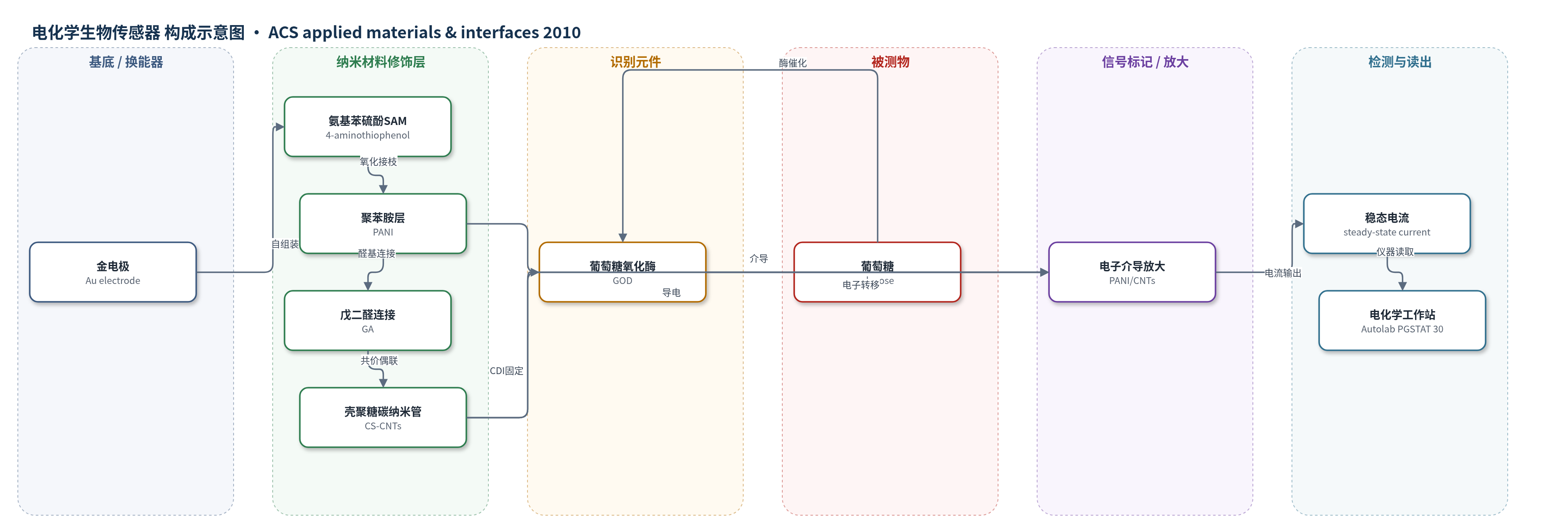
点击图片查看大图 · 依据论文自动绘制
传感器类型
电化学生物传感器
检测对象
葡萄糖(glucose);样品基质:磷酸盐缓冲液(PBS, pH 6.0),面向血液/体内血糖监测
检测原理
该传感器以金电极为换能基底,PANI与CS-CNTs构成导电/介导网络,GOD作为识别与催化元件。葡萄糖进入电极表面后,被GOD特异性催化氧化,其催化电子经CS-CNTs与PANI介导传递至金电极;碳纳米管提供导电通道,壳聚糖提供生物相容微环境,PANI作为导电聚合物促进电子转移。三维CS-CNTs网络增加GOD负载量并改善电极-酶接触,使稳态安培电流随葡萄糖浓度升高而增大。在0.5 V恒定电位下,1–20 mM范围内电流与浓度呈线性关系,体现酶催化-电子介导-安培读出机制。
检测灵敏度
LOD: about 0.1 mM;线性范围: 1-20 mM;灵敏度: 21 µA/(mM·cm2) (or 16.5 µA/mM)
效应效果
该传感器在0.5 V下响应时间约8–10 s,线性范围1–20 mM,覆盖体内监测常用5–20 mM。与无PANI电极相比,灵敏度由约5.2 µA/(mM·cm2)提高到21 µA/(mM·cm2),表观Michaelis-Menten常数由14.85 mM降至5.35 mM,酶活性更高。10个电极RSD约5.0%;4 °C PBS储存1个月和2个月分别保持约95%和83%初始电流。0.3 mM抗坏血酸和尿酸在1.0 mM葡萄糖下干扰可接受,对乙酰氨基酚干扰可忽略。共价固定可防止GOD流失,适用于高灵敏度葡萄糖传感。
传感器的构成
- 基底/换能器电极:金电极(Au electrode),提供导电基底与安培换能
- 自组装单分子层:4-氨基苯硫酚(4-aminothiophenol)SAM,提供氨基锚定位点
- 导电聚合物修饰层:聚苯胺(PANI),氧化接枝形成,介导电子转移并放大信号
- 双功能连接层:戊二醛(glutaraldehyde, GA),与PANI氨基反应并连接CS-CNTs
- 纳米复合材料修饰层:壳聚糖-偶联碳纳米管(CS-CNTs),由多壁碳纳米管(MWCNT)与壳聚糖共价偶联,形成三维导电网络
- 酶识别/催化元件:葡萄糖氧化酶(glucose oxidase, GOD),识别并催化葡萄糖
- 酶固定连接剂:1,4-碳酰二咪唑(1,4-carbonyldiimidazole, CDI),活化壳聚糖羟基并共价固定GOD
中文摘要
本文报道了一种基于共价固定壳聚糖-偶联碳纳米管(CS-CNTs)于聚苯胺(PANI)修饰金电极的安培型葡萄糖生物传感器(Au-g-PANI-c-(CS-CNTs)-GOD)。首先以4-氨基苯硫酚在金电极表面形成自组装单分子层,再通过氧化接枝聚合苯胺制备PANI层;随后以戊二醛为双功能连接剂,将CS-CNTs共价偶联到PANI修饰金基底上;最后以1,4-碳酰二咪唑为连接剂,将葡萄糖氧化酶(GOD)共价固定于壳聚糖侧链羟基上。XPS证实各功能化步骤,FESEM显示电极表面形成三维网络结构,可提供空间生物相容微环境,提高固定酶量与生物催化活性并促进电子转移。该传感器对葡萄糖在1–20 mM范围内呈线性响应,灵敏度为21 µA/(mM·cm2),重现性良好,储存2个月后仍保持初始响应电流的80%以上。
英文摘要
An amperometric glucose biosensor was prepared using polyaniline (PANI) and chitosan-coupled carbon nanotubes (CS-CNTs) as the signal amplifiers and glucose oxidase (GOD) as the glucose detector on a gold electrode (the Au-g-PANI-c-(CS-CNTs)-GOD biosensor). The PANI layer was prepared via oxidative graft polymerization of aniline from the gold electrode surface premodified by self-assembled monolayer of 4-aminothiophenol. CS-CNTs were covalently coupled to the PANI-modified gold substrate using glutaradehyde as a bifunctional linker. GOD was then covalently bonded to the pendant hydroxyl groups of chitosan using 1,4-carbonyldiimidazole as the bifunctional linker. The surface functionalization processes were ascertained by X-ray photoelectron spectroscopy (XPS) analyses. The field emission scanning electron microscopy (FESEM) images of the Au-g-PANI-c-(CS-CNTs) electrode revealed the formation of a three-dimensional surface network structure. The electrode could thus provide a more spatially biocompatible microenvironment to enhance the amount and biocatalytic activity of the immobilized enzyme and to better mediate the electron transfer. The resulting Au-g-PANI-c-(CS-CNTs)-GOD biosensor exhibited a linear response to glucose in the concentration range of 1-20 mM, good sensitivity (21 μA/(mM·cm(2))), good reproducibility, and retention of >80% of the initial response current after 2 months of storage.