组成图示
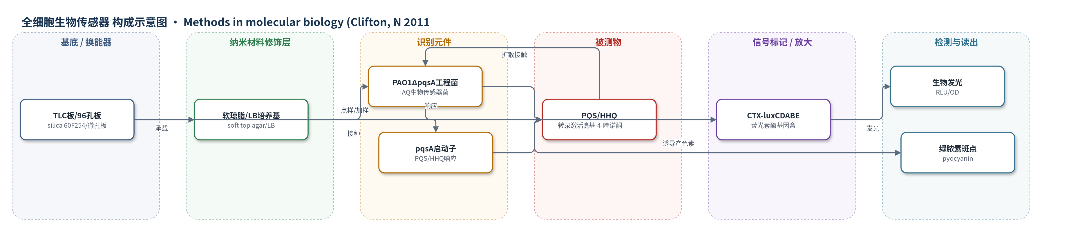
点击图片查看大图 · 依据论文自动绘制
传感器类型
全细胞生物传感器
检测对象
2-烷基-4-喹诺酮(2-alkyl-4-quinolones, AQs;含PQS、HHQ);样品基质:细菌培养上清、细胞有机溶剂提取物、TLC板斑点样品
检测原理
该检测基于全细胞生物报告菌。样品中的PQS或HHQ扩散至PAO1ΔpqsA CTX-lux::pqsA工程菌,激活pqsA启动子,驱动CTX-luxCDABE荧光素酶基因转录与翻译。荧光素酶催化荧光素氧化产生光子,形成生物发光;发光强度以RLU/OD表示,随AQ浓度升高而增强。TLC法中,AQ先在硅胶板上分离,覆盖含生物传感器的软琼脂后,AQ从斑点局部扩散,诱导对应位置发光及绿脓素绿色斑点。微孔板法中,样品与生物传感器共培养,96孔发光仪连续监测发光。基因表达与酶促发光提供信号放大,使方法适合批量筛选。
检测灵敏度
原文未报告检出限、线性范围、灵敏度斜率或相关系数。
效应效果
方法以生物发光和绿脓素斑点作为读出,可在复杂细菌上清中识别AQ,较UV-TLC对HHQ等弱荧光AQ更具判别性。文中以PAO1、PAO1ΔpqsH和PAO1ΔpqsA作为阳性、PQS阴性与AQ阴性对照,显示PAO1中PQS和HHQ斑点发光,PAO1ΔpqsH仅HHQ发光,pqsA突变体无发光。微孔板法中发光与AQ含量成正比,可用PQS/HHQ标准或PAO1上清作阳性对照。作者强调方法简单、无需昂贵精密仪器,适合大量细菌样品筛选;未报告LOD、线性范围、RSD或回收率。
传感器的构成
- 基底/载体:silica 60F254 TLC 板或 96-well 黑色透明底微孔板,承载样品与生物传感器
- 修饰/培养层:soft top agar 或 LB 培养基,覆盖 TLC 板或用于微孔板培养,支持生物传感器生长
- 生物传感细胞:Pseudomonas aeruginosa PAO1ΔpqsA 工程菌,缺失 pqsA,自身不产 AQ,作为识别与信号转导单元
- 识别元件:pqsA 启动子,响应 PQS/HHQ 并驱动报告基因表达
- 信号标记物:CTX-luxCDABE 荧光素酶基因盒,AQ 诱导后产生生物发光
- 样品基质:细菌培养上清或甲醇/乙酸乙酯提取物,提供 PQS/HHQ
- 读出介质:luminograph photon video camera 或 96-well luminometer,检测 RLU/OD 与绿脓素
中文摘要
2-烷基-4-喹诺酮(AQs)如2-庚基-3-羟基-4-喹诺酮(PQS)和2-庚基-4-喹诺酮(HHQ)是铜绿假单胞菌群体感应信号分子。本文介绍两种基于lux型铜绿假单胞菌AQ生物传感器菌株的AQ检测与定量方法。薄层色谱法中,将细菌细胞或培养上清的有机溶剂提取物点样于硅胶TLC板,展开干燥后覆盖含AQ生物传感器的软琼脂;生物报告菌受AQ诱导后,在对应位置产生生物发光和绿色绿脓素斑点。微孔板法中,将培养上清或提取物加入含AQ生物传感器的培养基,报告菌发光强度与样品AQ含量成正比。两种方法操作简便,无需昂贵精密仪器,适合大批量细菌样品筛选。
英文摘要
2-alkyl-4-quinolones (AQs) such as 2-heptyl-3-hydroxy-4-quinolone (PQS) and 2-heptyl-4-quinolone (HHQ) are quorum sensing signal molecules. Here we describe two methods for AQ detection and quantification that employ thin layer chromatography (TLC) and microtitre plate assays in combination with a lux-based Pseudomonas aeruginosa AQ biosensor strain. For TLC detection, organic solvent extracts of bacterial cells or spent culture supernatants are chromatographed on TLC plates, which are then dried and overlaid with the AQ biosensor. After detection by the bioreporter, AQs appear as both luminescent and green (pyocyanin) spots. For the microtitre assay, either spent bacterial culture supernatants or extracts are added to a growth medium containing the AQ biosensor. Light output by the bioreporter is proportional to the AQ content of the sample. The assays described are simple to perform, do not require sophisticated instrumentation, and are highly amenable to screening large numbers of bacterial samples.