组成图示
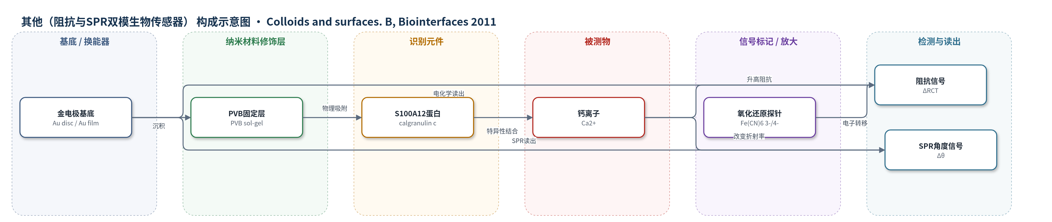
点击图片查看大图 · 依据论文自动绘制
传感器类型
其他(阻抗与SPR双模生物传感器)
检测对象
钙离子(Ca2+),样品基质为 PBS 缓冲液(pH 7.4)/去离子水,作者展望人血清
检测原理
该传感器基于猪 S100A12 蛋白对 Ca2+ 的特异性结合。S100A12 含 EF-hand 结构域,在 PVB 修饰的金电极表面物理吸附后仍保留钙结合活性。当 Ca2+ 与固定蛋白结合时,界面形成更致密的蛋白-钙复合物层,阻碍 [Fe(CN)6]3-/4- 氧化还原探针向金电极表面的扩散和电子转移,使 EIS 电荷转移电阻 RCT 增大、CV 峰电流降低;同时界面质量/折射率增加,使 SPR 共振角发生正偏移。Ca2+ 浓度越高,界面阻挡和 SPR 角偏移越大,在 12.5–200 mM 范围内呈线性响应。EDTA 螯合 Ca2+ 后 RCT 下降、SPR 角回落,证明结合可逆。该过程无需标记,直接以阻抗和 SPR 信号反映钙离子浓度。
检测灵敏度
线性范围: 12.5–200 mM
效应效果
该传感器对 Ca2+ 具有明显选择性:暴露于 Mg2+ 时 RCT 仅升至 3.72 kΩ,ΔRCT% 为 16.0;而 Ca2+ 使 RCT 升至约 11.0 kΩ,ΔRCT% 达 149.0。SPR 中非特异性离子无明显响应,Mg2+ 不改变光学信号。EDTA 螯合 Ca2+ 后 EIS 和 SPR 信号均回落,表明结合可逆。PVB 注入的变异系数为 10.87%,S100A12 吸附三次重复的 CV 为 7.23%,显示一定重现性。传感器在 12.5–200 mM 范围内线性响应,操作简便,作者认为其可作为可重复使用、高灵敏度的钙离子检测平台,并有望用于人血清钙测定,但文中未报告实际血清样品回收率或与 ELISA/HPLC/qPCR 等方法的直接对比。
传感器的构成
- 工作电极基底:金圆盘电极(Au,d=2 mm),提供电子转导与 SPR 金属膜基底
- 修饰层:聚乙烯醇丁醛(PVB,2% v/v 乙醇溶液),溶胶-凝胶法沉积,形成绝缘/固定基质并阻碍电子转移
- 识别元件:猪 S100A12 蛋白(calgranulin c,200 μg/mL),物理吸附于 PVB 层,通过 EF-hand 结合 Ca2+
- 氧化还原探针:K3[Fe(CN)6]/K4[Fe(CN)6](1:1,10 mM),用于 CV/EIS 监测界面电荷转移
- 参比电极:Ag/AgCl(饱和 KCl),提供稳定电位基准
- 辅助电极:铂丝(Pt),构成三电极体系
- 光学换能器:金镀 SPR 传感表面(Au-PVB-S100A12),无标记检测界面质量/折射率变化
- 验证试剂:EDTA,螯合 Ca2+ 验证结合可逆
中文摘要
本文报道了一种无标记钙离子生物传感器的构建与表征。作者将猪源 S100A12 蛋白(calgranulin c)与聚乙烯醇丁醛(PVB)混合后,通过溶胶-凝胶法固定在金电极表面,形成 Au-PVB-S100A12 修饰电极。采用循环伏安法(CV)、电化学阻抗谱(EIS)、扫描电镜(SEM)和表面等离子共振(SPR)对电极进行表征。SEM 显示 PVB 在金表面呈球状异质分布;CV 和 EIS 表明 PVB-S100A12 层部分阻碍了 [Fe(CN)6]3-/4- 氧化还原探针的电子转移,且钙离子结合后界面电荷转移阻力进一步升高,证实固定蛋白对 Ca2+ 具有特异性响应。该传感器在 12.5–200 mM Ca2+ 范围内呈宽线性响应。SPR 结果显示,PVB 上吸附 S100A12 后共振角增加 1184.32 m°,相当于约 9.84 ng/mm2 蛋白吸附量;Ca2+ 结合后再增加 581.66 m°,而非特异性离子无明显响应。作者认为该平台有望用于可重复使用、高灵敏度的临床钙离子检测。
英文摘要
We describe the development of a label free method to analyze the interactions between Ca(2+) and the porcine S100A12 protein immobilized on polyvinyl butyral (PVB). The modified gold electrodes were characterized using cyclic voltammetry (CV), electrochemical impedance spectroscopy (EIS), scanning electron microscopy (SEM) and surface plasmon resonance (SPR) techniques. SEM analyses of PVB and PVB-S100A12 showed a heterogeneous distribution of PVB spherules on gold surface. EIS and CV measurements have shown that redox probe reactions on the modified gold electrodes were partially blocked due the adsorption of PVB-S100A12, and confirm the existence of a positive response of the immobilized S100A12 to the presence of calcium ions. The biosensor exhibited a wide linear response to Ca(2+) concentrations ranging from 12.5 to 200mM. The PVB-S100A12 seems to be bound to the gold electrode surface by physical adsorption; we observed an increase of 1184.32m° in the SPR angle after the adsorption of the protein on the PVB surface (in an indication that 9.84ng of S100A12 are adsorbed per mm(2) of the Au-PVB electrode), followed by a further increase of 581.66m° after attachment of the Ca(2+) ions. In addition, no SPR response is obtained for non-specific ions. These studies might be useful as a platform for the design of new reusable and sensitive biosensing devices that could find use in the clinical applications.