组成图示
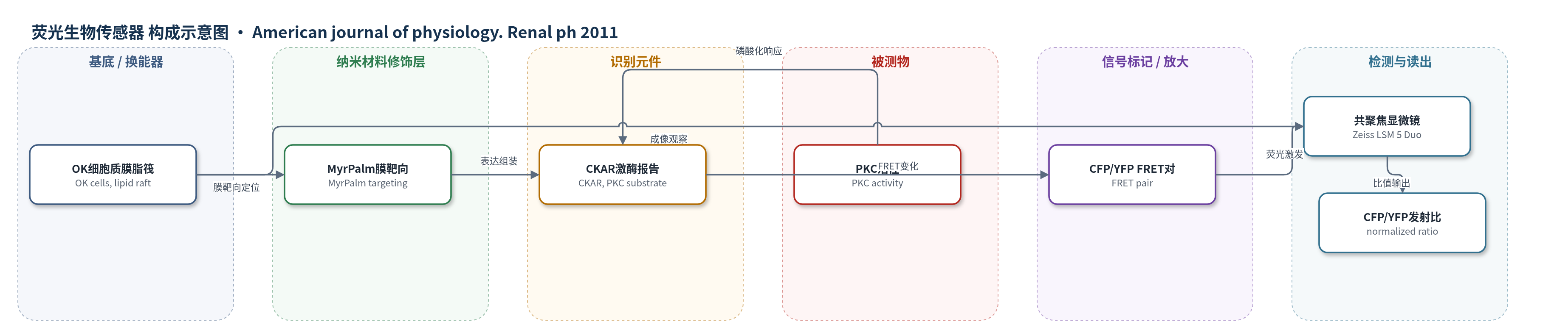
点击图片查看大图 · 依据论文自动绘制
传感器类型
荧光生物传感器
检测对象
蛋白激酶C活性(protein kinase C, PKC activity);样品基质:活体袋狸肾细胞(OK cells)质膜/脂筏。
检测原理
该检测基于靶向质膜脂筏的FRET激酶活性报告蛋白MyrPalm-CKAR。PTH与OK细胞PTH1受体结合后激活蛋白激酶C(PKC),PKC磷酸化CKAR报告元件,改变CFP/YFP荧光共振能量转移(FRET)效率。共聚焦显微镜分别以488 nm和561 nm激光激发CFP/YFP,测量归一化CFP/YFP发射比;PKC活性升高时FRET比发生可测变化,从而在活细胞中实时反映PTH诱导的PKC激活时间过程。该策略无需固定或裂解细胞,可直接在质膜/脂筏界面监测激酶活性。
检测灵敏度
原文未报告LOD、线性范围、灵敏度斜率或相关系数。
效应效果
在活体OK细胞中,PTH(10^-7 M)处理后60–90 s内PKC即达到最大激活。GFP-Npt2a/mCherry-NHERF-1荧光比在10 min开始下降,20 min出现显著变化,30 min达到最大,半时约15–20 min;NHERF-1相对丰度基本不变。GFP-ezrin/mCherry-NHERF-1比值未受PTH影响,共免疫沉淀显示ezrin/NHERF-1比值控制组0.17±0.01、PTH组0.18±0.01,反向沉淀为0.54±0.05与0.52±0.07,差异无统计学意义。结果表明该FRET报告系统可区分PTH对Npt2a/NHERF-1与ezrin/NHERF-1复合物的选择性作用。
传感器的构成
- 基底/换能界面:活体袋狸肾细胞(OK cells)质膜/脂筏区域,作为FRET报告蛋白表达与定位的细胞界面。
- 膜靶向修饰层:MyrPalm序列,将激酶活性报告蛋白CKAR靶向至质膜脂筏区域。
- 识别/响应元件:CKAR(kinase activity reporter),作为PKC活性响应元件,接受PKC磷酸化。
- 信号标记物:CFP/YFP FRET荧光蛋白对(cyan/yellow emission),PKC激活引起FRET效率变化。
- 读出系统:Zeiss LSM 5 Duo共聚焦显微镜,488 nm Ar激光与561 nm二极管激光激发,MetaMorph软件计算CFP/YFP归一化发射比。
中文摘要
甲状旁腺激素(PTH)通过破坏钠依赖性磷酸盐转运体2A(Npt2a)与钠-氢交换调节因子1(NHERF-1)的结合,抑制肾近端小管磷酸盐重吸收,该过程由蛋白激酶C(PKC)激活启动。为阐明动态事件顺序,作者在活体袋狸肾细胞(OK cells)中研究其时间过程。利用FRET生物传感器发现,PTH可在数秒至数分钟内激活细胞内PKC。在表达GFP-Npt2a和mCherry-NHERF的细胞中,PTH不影响NHERF-1相对丰度,但Npt2a/NHERF-1比值显著且随时间下降,最大解离半时约15–20 min。相比之下,PTH对顶膜处GFP-ezrin与mCherry-NHERF-1荧光比无影响。结果表明,PTH快速激活PKC并导致Npt2a/NHERF-1复合物解离;NHERF-1与ezrin的结合及顶膜定位未受扰动,有利于激素反应终止后Npt2a及其他NHERF-1靶蛋白快速募集和膜再插入。
英文摘要
Parathyroid hormone (PTH) inhibits the reabsorption of phosphate in the renal proximal tubule by disrupting the binding of the sodium-dependent phosphate transporter 2A (Npt2a) to the adapter protein sodium-hydrogen exchanger regulatory factor-1 (NHERF-1), a process initiated by activation of protein kinase C (PKC). To gain additional insights into the dynamic sequence of events, the time course of these responses was studied in living opossum kidney (OK) cells. Using a FRET-based biosensor, we found that PTH activated intracellular PKC within seconds to minutes. In cells expressing GFP-Npt2a and mCherry-NHERF, PTH did not affect the relative abundance of NHERF-1 but there was a significant and time-dependent decrease in the Npt2a/NHERF-1 ratio. The half-time to maximal dissociation was 15 to 20 min. By contrast, PTH had no effect on the fluorescence ratio for GFP-ezrin compared with mCherry-NHERF-1 at the apical surface. These experiments establish that PTH treatment of proximal tubule OK cells leads to rapid activation of PKC with the subsequent dissociation of Npt2a/NHERF-1 complexes. The association of NHERF-1 with Ezrin and their localization at the apical membrane, however, was unperturbed by PTH, thereby enabling the rapid recruitment and membrane reinsertion of Npt2a and other NHERF-1 targets on termination of the hormone response.