组成图示
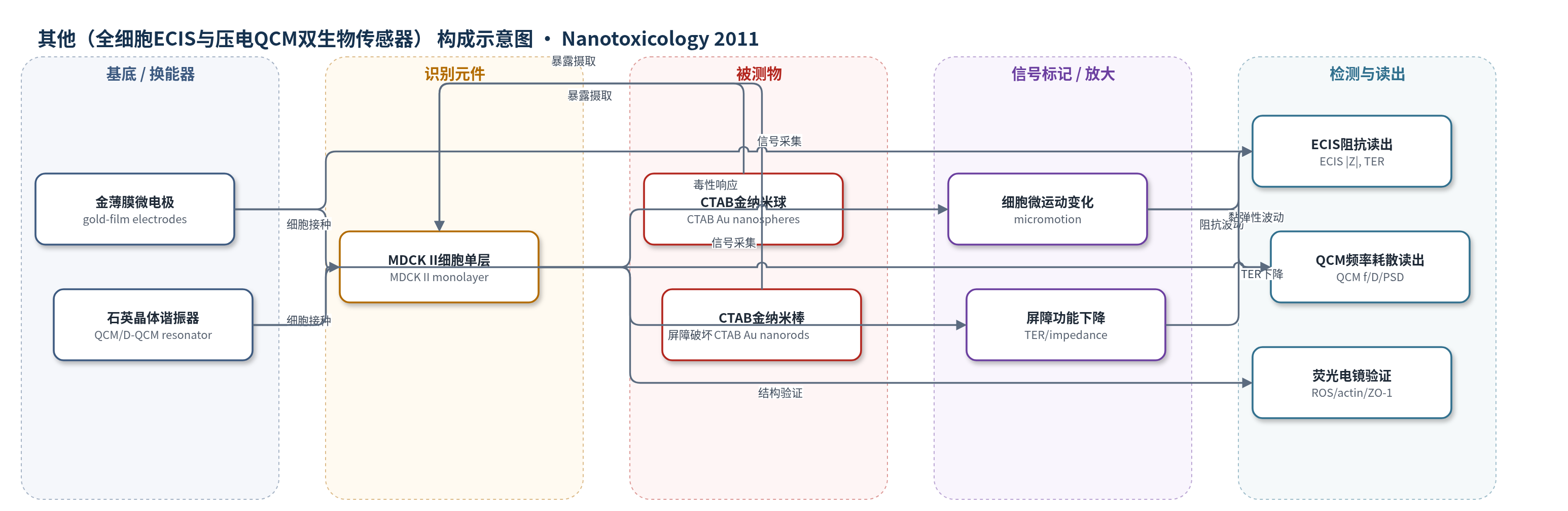
点击图片查看大图 · 依据论文自动绘制
传感器类型
其他(全细胞ECIS与压电QCM双生物传感器)
检测对象
CTAB包覆金纳米球(CTAB-coated Au nanospheres)、CTAB包覆金纳米棒(CTAB-coated Au nanorods);样品基质为MDCK II上皮细胞培养液/细胞单层。
检测原理
本方法以MDCK II上皮细胞单层作为全细胞识别/响应元件。CTAB包覆金纳米颗粒加入培养液后,经巨胞饮进入细胞,在早期内体/多泡体中聚集;球形颗粒因体积和表面积更大,聚集时更易释放表面CTAB,诱发ROS、细胞骨架破坏和细胞间接触解体。ECIS中,细胞体近似绝缘体,改变金微电极周围电流路径,细胞形状波动和屏障完整性表现为4 kHz阻抗|Z|、同相电压波动及TER;毒性增强时微运动减弱、|Z|和TER下降。D-QCM中,细胞黏弹性负载改变石英晶体剪切振荡的频率f与耗散D,FFT/PSD低频斜率反映细胞活力。信号随纳米颗粒浓度升高而降低,球形颗粒响应强于棒形。
检测灵敏度
原文未报告LOD、线性范围、灵敏度斜率或相关系数;报告IC50(μg/mL,平均标准差±0.1 MTS、±0.15 micromotionECIS、±0.5 micromotionF-QCM,n=3):MTS s-GNPs 24 h 0.3、r-GNPs 48 h 1.8;Micromotion ECIS s-GNPs 24 h 0.2、r-GNPs 24 h 16.3、s-GNPs 48 h 0.1、r-GNPs 48 h 1.1;Micromotion F-QCM s-GNPs 24 h 1.5、r-GNPs 24 h 16.0、s-GNPs 48 h 2.3、r-GNPs 48 h 12.0。
效应效果
两种无标记传感器独立给出一致结果:球形CTAB金纳米颗粒毒性显著高于棒形。9 μg/mL球形颗粒使ECIS阻抗|Z|下降60–70%,TER在24 h内不可逆丧失;棒形颗粒基本不影响屏障功能。ECIS微运动比MTS更敏感,F-QCM在低浓度下响应较弱。IC50显示球形颗粒24 h ECIS为0.2 μg/mL、48 h为0.1 μg/mL,棒形24 h为16.3 μg/mL、48 h为1.1 μg/mL。15 μg/mL暴露后每细胞约2000–3000颗粒,聚集体约48±18(棒)或44±12(球)颗粒/聚集体。作者认为ECIS与D-QCM组合可实时、非侵入监测纳米毒性,适合高通量筛查。
传感器的构成
- 换能器电极:金薄膜微电极阵列(gold-film microelectrodes, 8W1E),ECIS中承载MDCK II细胞并采集交流阻抗。
- 压电换能器:石英晶体谐振器(quartz crystal resonator, QCM/D-QCM),检测细胞黏弹性引起的频率与耗散变化。
- 识别/响应元件:MDCK II上皮细胞单层(MDCK II cell monolayer),作为全细胞响应层,其形状、黏弹性和细胞间接触随金纳米颗粒暴露改变。
- 读出电路:1 V交流信号、1 MΩ串联电阻、4 kHz阻抗采集(ECIS);频率/耗散采集与FFT/PSD分析(D-QCM)。
- 培养腔:特氟龙腔(teflon-chambers)与37℃、5% CO2培养环境,维持QCM细胞活性。
中文摘要
本研究采用两种无标记、非侵入式生物传感器装置监测纳米颗粒暴露:石英晶体微天平(QCM)检测细胞形状与黏弹性,电细胞-基底阻抗传感(ECIS)检测细胞运动及上皮细胞间接触动力学。作者合成并研究了两种不同形状、表面均包覆十六烷基三甲基溴化铵(CTAB)的金纳米颗粒:直径约43±4 nm的球形颗粒和约38±7 nm×17±3 nm的棒状颗粒。剂量-响应实验结合常规细胞毒性检测、荧光显微镜和暗场显微镜,以观察颗粒在细胞内的分布。结果表明,在相同表面功能化条件下,球形金纳米颗粒通常比棒状颗粒毒性更高,且被细胞摄取更高效。作者将CTAB包覆球形颗粒毒性高于棒状颗粒主要归因于颗粒在细胞内聚集后释放更多有毒CTAB。
英文摘要
Nanoparticle exposure is monitored by a combination of two label-free and non-invasive biosensor devices which detect cellular shape and viscoelasticity (quartz crystal microbalance), cell motility and the dynamics of epithelial cell-cell contacts (electric cell-substrate impedance sensing). With these tools we have studied the impact of nanoparticle shape on cellular physiology. Gold (Au) nanoparticles coated with CTAB were synthesized and studied in two distinct shapes: Spheres with a diameter of (43 ± 4) nm and rods with a size of (38 ± 7) nm × (17 ± 3) nm. Dose-response experiments were accompanied by conventional cytotoxicity tests as well as fluorescence and dark-field microscopy to visualize the intracellular particle distribution. We found that spherical gold nanoparticles with identical surface functionalization are generally more toxic and more efficiently ingested than rod-shaped particles. We largely attribute the higher toxicity of CTAB-coated spheres as compared to rod-shaped particles to a higher release of toxic CTAB upon intracellular aggregation.