组成图示
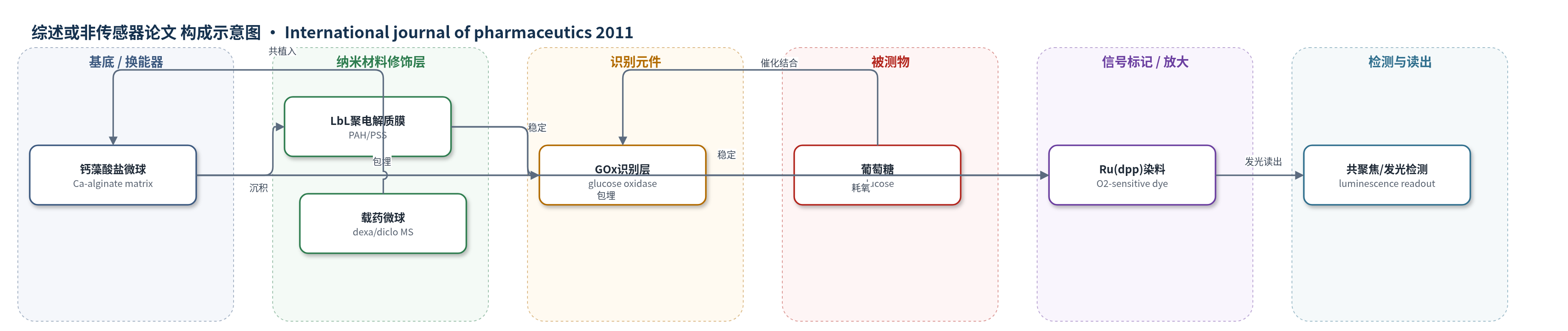
点击图片查看大图 · 依据论文自动绘制
传感器类型
综述或非传感器论文
检测对象
葡萄糖(glucose);样品基质:皮下组织间液/模拟组织间液(SIF)
检测原理
该模型“智能纹身”葡萄糖传感器以钙藻酸盐微球为基质,包埋葡萄糖氧化酶(GOx)和氧敏感发光染料 Ru(dpp)。葡萄糖扩散进入微球后,GOx 催化葡萄糖氧化并消耗局部氧;Ru(dpp) 的发光强度或寿命随氧浓度变化,从而将葡萄糖浓度转换为光学信号。LbL 聚电解质膜(PAH/PSS)用于稳定酶与染料、控制物质传输并降低突释。载药微球共植入后局部释放地塞米松或双氯芬酸钠,抑制植入部位炎症与纤维化,以维持传感器界面通透性。本文未报告该传感器的定量检测性能。
检测灵敏度
—
效应效果
本文未报告葡萄糖传感器的定量分析性能。药物载体方面,载药微球包封率约77±8%;通过未包衣与PAH/PSS包衣微球配比,可在30天实现约100%零级释放,SIF中30天累计释放为96%(地塞米松)和97%(双氯芬酸)。L929细胞活力均>80%。体内SD大鼠H&E计数显示:空白微球第7天中性粒细胞70±5/HPF,第28天形成厚纤维囊;地塞米松组40±4/HPF、双氯芬酸组45±7/HPF且无纤维囊;葡萄糖传感器组100±5/HPF并形成厚纤维囊,与地塞米松/双氯芬酸共植入后分别降至41±5和52±7/HPF且无纤维囊,提示可改善植入界面炎症。
传感器的构成
- 基底/载体:海藻酸钠(sodium alginate)微球,经氯化钙(CaCl2)交联形成钙藻酸盐(calcium alginate)微球,作为酶与染料包埋基质
- 识别元件:葡萄糖氧化酶(GOx,Aspergillus niger Type VII),催化葡萄糖氧化并消耗氧
- 信号标记物:氧敏感发光染料 Ru(dpp)(ruthenium-tris(4,7-diphenyl-1,10-phenanthroline) dichloride),用于光学/发光读出
- 修饰层:聚电解质层层自组装(LbL)纳米薄膜,由聚(4-苯乙烯磺酸钠)(PSS)与聚(烯丙基胺盐酸盐)(PAH,FITC标记)交替沉积,稳定酶/染料并控制传输
- 抗炎载体:载地塞米松(dexamethasone)或双氯芬酸钠(diclofenac sodium)的钙藻酸盐微球,局部释放抗炎药
- 涂层:PAH/PSS 聚电解质涂层,降低药物突释并延长零级释放
中文摘要
本研究旨在开发纳米工程化海藻酸微球,用于局部递送抗炎药物(地塞米松和双氯芬酸钠),以改善用于连续血糖监测的植入式“智能纹身”葡萄糖生物传感器的生物相容性。作者制备并表征了未包衣和聚电解质包衣微球的体外药物释放行为,并用L929细胞系评价生物相容性;随后在Sprague–Dawley大鼠中进行初步体内研究,通过苏木精–伊红染色分析植入相关炎症与纤维化。载药微球可在30天内以零级动力学受控释放药物,层层自组装技术有效降低突释。细胞实验显示材料无细胞毒性,所有样品细胞活力均>80%。体内结果表明两种药物均可抑制植入部位炎症,控制植入/组织界面。载药载体与植入生物传感器联合使用有望提高传感器生物相容性与功能,提示海藻酸微球在“智能纹身”葡萄糖传感器中的应用潜力。
英文摘要
The aim of this research was to develop nanoengineered alginate microspheres for localized delivery of anti-inflammatory drugs (dexamethasone and diclofenac sodium) for implantable "Smart tattoo" glucose biosensor used for continuous glucose monitoring. The formulation was prepared and characterized for in vitro drug release from uncoated and polyelectrolyte-coated microparticles. Biocompatibility was then tested using L929 cell-line; pilot in vivo studies with Sprague-Dawley (SD) rat subjects were performed to test the suppression of inflammation and fibrosis associated with implantation and was analyzed using standard hematoxylin and eosin staining method. The drug-loaded microspheres were able to deliver the drug for 30 days at a controlled rate with zero-order kinetics. The layer-by-layer self-assembly technique was used to effectively limit the burst release of drug from the matrix. Cell culture studies prove that the material are not cytotoxic and showed acceptable >80% cell viability in all the tested samples. In vivo studies show that both drugs were successful in controlling the implant/tissue interface by suppressing inflammation at the implant site. It was clearly evident that the combined approach of drug loaded carriers along with implanted biosensor shows promise in improving sensor biocompatibility and functionality. Thus, suggesting potential application of alginate microspheres as "smart-tattoo" glucose sensors.