组成图示
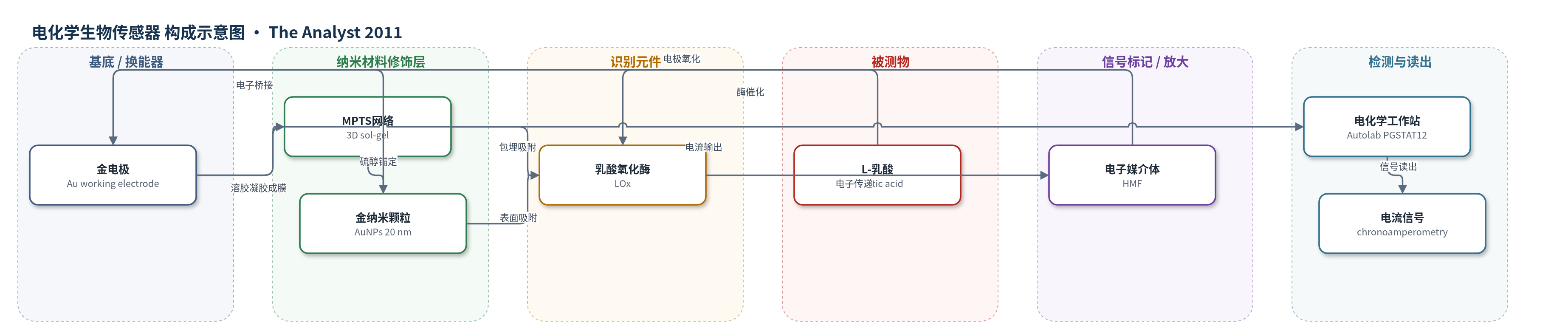
点击图片查看大图 · 依据论文自动绘制
传感器类型
电化学生物传感器
检测对象
L-乳酸(L-lactic acid / lactate);样品基质:磷酸盐缓冲液(PBS,pH 7.0),文中提及可应用于血液和食品样品
检测原理
传感器以LOx为识别元件,催化L-乳酸氧化为丙酮酸。传统反应以O2为电子受体生成H2O2,本工作用可溶性媒介体HMF替代O2作为人工电子受体。LOx催化过程中产生的电子经HMF传递至金电极,HMF在+0.30 V被氧化,产生与乳酸浓度成正比的计时电流。MPTS三维溶胶-凝胶网络包埋LOx,保持其催化活性并提高酶负载量;AuNPs分散在网络中作为微小电子传导中心,缩短电子转移距离并增强电流。因此乳酸浓度越高,催化电流越大,通过电化学工作站读取电流即可定量。
检测灵敏度
LOD: 4.0 mM;线性范围: 50 mM–0.25 mM;灵敏度: 3.4 mA mM^-1;r = 0.994
效应效果
与无AuNPs的Au/MPTS/LOx相比,AuNPs显著提高电流响应。重复性RSD为2%(同一传感器测0.1 mM乳酸10次),重现性RSD为5%(三个不同传感器)。4°C保存10天后信号衰减10%。干扰实验显示,0.1 mM酒石酸、柠檬酸、葡萄糖、果糖不改变响应;0.1 mM抗坏血酸有干扰,但10 mM无影响。QCM表明MPTS/AuNPs网络可负载619 ng cm^-2 LOx,约为裸金或DTSP金表面的3.5倍。作者认为该杂化网络适合乳酸检测,可应用于医学和食品工业。
传感器的构成
- 基底/换能器电极:多晶金电极(Au),经抛光和电化学活化,提供导电基底和硫醇锚定位点
- 溶胶-凝胶网络层:(3-巯基丙基)三甲氧基硅烷(MPTS)水解缩合形成的三维硅酸盐聚合物网络,提供生物相容包埋环境并含硫醇尾基
- 纳米材料修饰层:胶体金纳米颗粒(AuNPs,约20 nm),通过硫醇基化学吸附于MPTS网络,作为电子传导中心促进电子转移
- 识别元件:乳酸氧化酶(LOx),包埋/吸附于MPTS/AuNPs网络,催化L-乳酸氧化为丙酮酸,并将电子传递给HMF
- 电子媒介体:羟甲基环戊二烯铁茂(HMF),可溶性人工电子受体,在+0.30 V被电极氧化,传递LOx催化电子
- 检测介质:0.1 M磷酸盐缓冲液(PBS,pH 7.0),维持酶活性和电化学测量环境
中文摘要
本文设计并表征了一种新型纳米结构有机–无机杂化材料,并将其用于L-乳酸的电化学测定。该材料将乳酸氧化酶(LOx)和金纳米颗粒(AuNPs)整合到由(3-巯基丙基)三甲氧基硅烷(MPTS)在金表面形成的溶胶–凝胶三维聚合物网络中。MPTS可形成含大量硫醇尾基的三维网络,既能通过硫醇基锚定于金表面,又能结合AuNPs;同时提供生物相容环境,保持LOx催化活性并允许高负载量,从而提高传感器灵敏度。作者采用石英晶体微天平(QCM)、扫描电子显微镜(SEM)和原子力显微镜(AFM)表征该平台,获得LOx吸附动力学、酶负载量和纳米形貌信息。最终传感器对L-乳酸在50 mM–0.25 mM范围内呈线性响应,灵敏度为3.4 mA mM^-1,检出限为4.0 mM。
英文摘要
The design and characterization of a new nanostructured organic-inorganic hybrid material and its application to L-lactic acid determination are described. This material is based on the integration of the enzyme lactate oxidase (LOx) and gold nanoparticles (AuNPs) into a sol-gel 3D polymeric network derived from (3-mercaptopropyl)-trimethoxysilane (MPTS) previously formed onto a gold surface. MPTS presents the advantage of forming a 3D polymeric network containing a large number of thiol tail groups distributed throughout its structure that enable both its anchoring onto gold surfaces and the AuNPs incorporation. Moreover, this matrix provides a biocompatible environment that preserves the catalytic activity of LOx after its immobilization and allows the incorporation of a high amount of enzyme, which is expected to improve the sensitivity of the final biosensing device. Characterization of the designed biosensing platform was performed using quartz crystal microbalance (QCM), scanning electron microscopy (SEM) and atomic force microscopy (AFM) techniques. From the conjunction of these techniques, information about (i) the kinetic of LOx adsorption process in real time, (ii) the amount of LOx incorporated into the network, and (iii) the morphological characteristics at the nanometre level of the designed biosensing material was obtained. This information is very useful on the development of successful biosensing devices. Finally, the response of the biosensor to L-lactic acid was evaluated. The biosensor responds linearly to L-lactic acid in the range of 50 µM to 0.25 mM, with a sensitivity of 3.4 µA mM(-1) and a detection limit of 4.0 µM.