组成图示
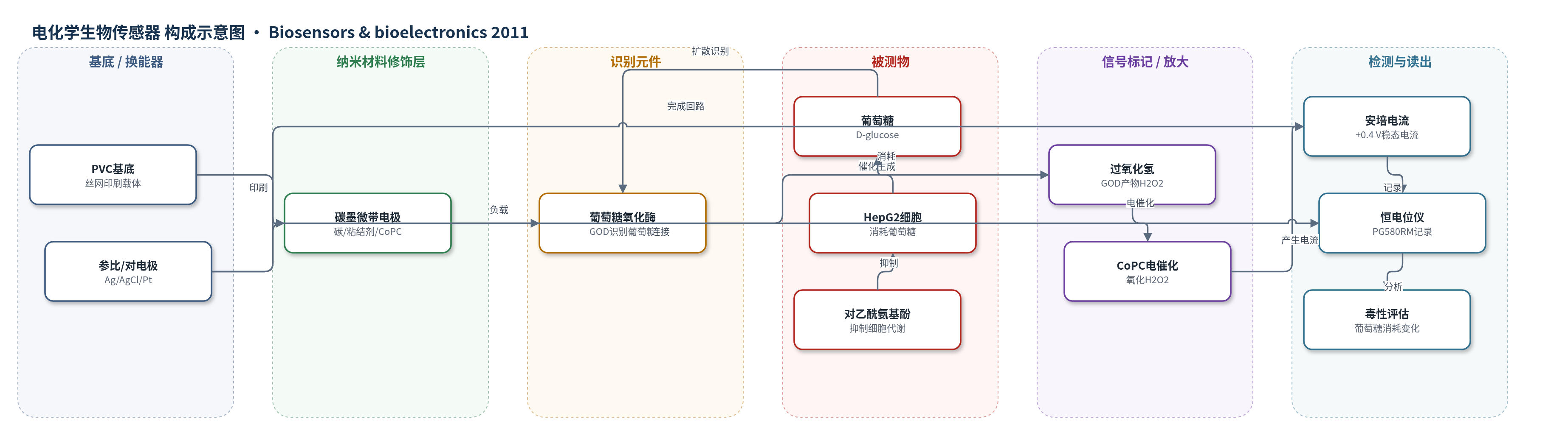
点击图片查看大图 · 依据论文自动绘制
传感器类型
电化学生物传感器
检测对象
葡萄糖(glucose, D-glucose)、对乙酰氨基酚(paracetamol,间接毒性评估);样品基质:磷酸盐缓冲液、DMEM/FBS细胞培养基、HepG2单层/球体培养液
检测原理
葡萄糖扩散至微带电极表面,被葡萄糖氧化酶(GOD)催化氧化,生成过氧化氢(H2O2)。在+0.4 V下,碳墨中的钴酞菁(CoPC)电催化氧化H2O2,产生与葡萄糖浓度成正比的阳极电流。微带电极在静止条件下以径向扩散为主,形成稳态安培响应,因此电流可连续反映培养基中葡萄糖浓度。HepG2细胞或球体代谢葡萄糖会使葡萄糖浓度下降,电流随之降低;对乙酰氨基酚抑制细胞葡萄糖摄取后,电流下降速率减缓,从而通过比较对照孔与处理孔的电流变化评估毒性。系统无额外信号放大,主要依赖酶催化与电催化转换。
检测灵敏度
线性范围: 0–10 mM(动态范围 30 mM);灵敏度: 7.84 nA mM−1(缓冲液,R2 = 0.957)、8.43 nA mM−1(培养基,R2 = 0.993);R2 = 0.981(与分光光度法)
效应效果
传感器24 h稳态响应稳定,缓冲液与L-15培养基校准一致(PLS R2=0.992);全培养基pH 7.3–8.4响应变化<5%。24 h内传感器仅消耗89 nmol葡萄糖,占24 mM起始浓度<0.15%。与分光光度法R2=0.981,至17 mM一致性较好但存在负偏差。重复孔CV从1 h的1.4%增至24 h的9.6%;球体电流变化CV为11%。HepG2单层电流下降速率与细胞密度线性相关(R2=0.985),24 h剩余葡萄糖为23.4–6.5 mM。对乙酰氨基酚1 mM时6 h抑制95%;0.5 mM时16 h抑制15%;三次实验抑制率为95%、81%、30%(CV 40%)。作者认为可发展为体外毒性测试平台。
传感器的构成
- 基底/换能器电极:PVC基底上丝网印刷水基碳墨工作电极(GEM C2041124D3),3 mm×3 mm、厚20 μm,切割暴露3 mm×20 μm微带边缘,提供导电微带换能器
- 电催化修饰层:碳墨中的碳、粘结剂和钴酞菁(CoPC),作为电催化剂氧化H2O2
- 识别元件:葡萄糖氧化酶(GOD),催化葡萄糖氧化生成H2O2
- 参比电极:Ag/AgCl墨水(GEM C61003P7)丝网印刷于0.5 mm热缩PVC上,2 mm宽条带,提供稳定参比电位
- 对电极:Pt线,完成三电极电化学回路
- 传感器头/样品室:自制聚丙烯三电极传感器头,推入12孔培养板,含1 mm孔和无菌滤头,固定电极并允许气体交换
- 检测仪器:PG580RM 5通道恒电位仪与UiEChem软件,施加+0.4 V并记录安培电流
中文摘要
本文报道了一种用于细胞培养毒性实时监测的丝网印刷微带葡萄糖生物传感器系统。该传感器由含钴酞菁(CoPC)氧化还原介质和葡萄糖氧化酶(GOD)的水基碳墨丝网印刷而成,在12孔组织培养板中于37 ℃以安培法(+0.4 V)连续运行。24 h内,稳态电流对葡萄糖浓度在0–10 mM范围内线性,动态范围约30 mM;与分光光度法相比,线性区相关系数R2为0.981。24 h内传感器仅消耗初始24 mM葡萄糖的约0.15%(89 nmol)。在全培养基中,pH 7.3–8.4范围内响应稳定。HepG2单层培养中,安培电流随葡萄糖被细胞代谢而下降,下降速率与细胞密度(0–1×10^6 cells/mL)相关;HepG2三维球体也能代谢葡萄糖,且速率在6–15天内基本不变。利用球体检测肝毒性物质对乙酰氨基酚:1 mM时6 h后葡萄糖摄取抑制约95%,500 μM时16 h后抑制约15%。作者认为该系统可发展为体外毒性测试平台。
英文摘要
Microband biosensors, screen-printed from a water-based carbon ink containing cobalt phthalocyanine redox mediator and glucose oxidase (GOD) enzyme, were used to monitor glucose levels continuously in buffer and culture medium. Five biosensors were operated amperometrically (E(app) of +0.4V), in a 12-well tissue culture plate system at 37°C, using a multipotentiostat. After 24 h, a linear calibration plot was obtained from steady-state current responses for glucose concentrations up to 10 mM (dynamic range 30 mM). Within the linear region, a correlation coefficient (R(2)) of 0.981 was obtained between biosensor and spectrophotometric assays. Over 24 h, an estimated 0.15% (89 nmol) of the starting glucose concentration (24 mM) was consumed by the microbiosensor. The sensitivity of the biosensor response in full culture medium was stable between pHs 7.3 and 8.4. Amperometric responses for HepG2 monolayer cultures decreased with time in inverse proportionality to cell number (for 0 to 10(6) cell/ml), as glucose was being metabolised. HepG2 3D cultures (spheroids) were also shown to metabolise glucose, at a rate which was independent of spheroid age (between 6 and 15 days). Spheroids were used to assay the effect of a typical hepatotoxin, paracetamol. At 1 mM paracetamol, glucose uptake was inhibited by 95% after 6 h in culture; at 500 μM, around 15% inhibition was observed after 16 h. This microband biosensor culture system could form the basis for an in vitro toxicity testing system.