组成图示
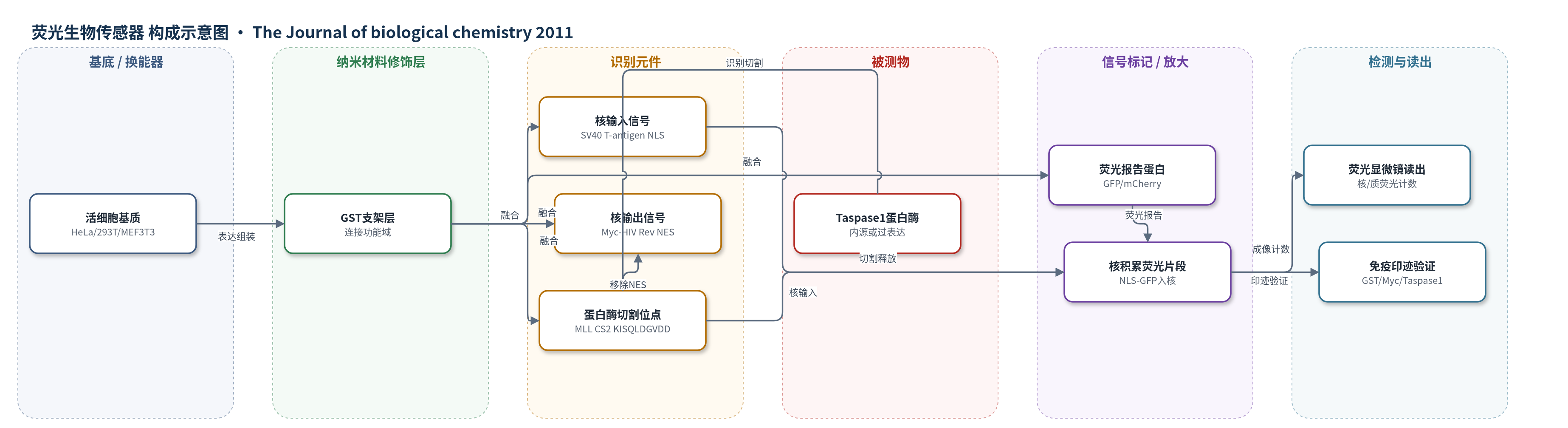
点击图片查看大图 · 依据论文自动绘制
传感器类型
荧光生物传感器
检测对象
Taspase1蛋白酶活性(Threonine Aspartase 1, Taspase1);样品基质:HeLa、293T、MEF3T3等活细胞及细胞裂解液
检测原理
BioTasp融合蛋白由NLS-GFP/GST-MLL CS2-NESRev组成,因核输入信号NLS与核输出信号NESRev共同作用,主要在细胞质中表达并持续核质穿梭。当细胞内存在活性Taspase1时,其以P1位天冬氨酸为核心的切割位点识别CS2,并在Asp后发生蛋白水解切割,移除Myc-NESRev。切割后NLS-GFP片段失去核输出能力,在细胞核中积累,形成核荧光信号。Taspase1活性越高,传感器被切割比例越高,核阳性细胞比例和核荧光强度越大;无活性TaspTV突变体或无关蛋白酶不能诱导核积累。该体系利用细胞内蛋白酶催化循环实现信号放大,并通过荧光显微镜计数或免疫印迹读取。
检测灵敏度
—
效应效果
选择性良好:无活性Taspase1突变体TaspTV/TaspTA、caspase3/caspase9及Rev-BFP均不诱导核积累;BioTaspmut和BioCasp3保持细胞质定位。1 μg BioTasp与0.1 μg Tasp-BFP共转染即可显著诱导切割和核积累,p<0.0001;至少200个细胞、三次独立实验。稳定诱导系BioTaspi可检测内源Taspase1。作者验证MLL、TF2A、USF2、MYO1F、NXF2/5切割位点,并证明全长TF2A-GFP和USF2-GFP可被切割,NXF2/5效率较低。该体系可用于高通量筛选Taspase1抑制剂及研究其病理功能。
传感器的构成
- 细胞基质:HeLa、293T、MEF3T3等活细胞,提供Taspase1表达、核质转运与蛋白酶活性环境
- 支架/连接层:GST,连接NLS-GFP与切割位点/NESRev,维持融合蛋白结构
- 荧光报告层:GFP或mCherry,提供可被荧光显微镜读取的荧光信号
- 核输入元件:SV40 large T-antigen NLS,驱动融合蛋白进入细胞核
- 核输出元件:Myc表位标记HIV-1 Rev NES(NESRev),维持融合蛋白主要位于细胞质并可被切割移除
- 识别/切割元件:Taspase1切割位点(MLL CS2:KISQLDGVDD及其变体),被Taspase1特异性水解
- 信号读出层:荧光显微镜、Hoechst 33258核染色及免疫印迹(anti-GST、anti-Myc、anti-Taspase1、anti-GFP),读取核积累或切割产物
中文摘要
Taspase1是一种负责切割细胞内底物的苏氨酸蛋白酶,其功能失调可能参与发育调控和疾病发生,但其完整底物谱及高效、底物特异性切割的生化要求尚未明确,且目前缺乏可用于该蛋白酶的细胞学检测体系。本研究开发了高效细胞内转位生物传感器体系,用于在活细胞中检测Taspase1的反式切割活性。该模块化传感器携带不同Taspase1切割位点变体,主要定位于细胞质;表达活性Taspase1可触发传感器蛋白水解切割并使其在细胞核中积累,而无活性突变体或无关蛋白酶不能诱导该信号。结合扫描突变分析,作者在液体和实体肿瘤细胞系中鉴定了Taspase1高效加工所需的序列与空间要求,并定义了改进的共识识别序列Q3(F/I/L/V)2D12G1-X2-D3-D4-,从而首次实现人源Taspase1降解组的基因组范围生物信息学鉴定。27个最可能底物包括细胞质和核蛋白,如USF2和NXF2/5。所选底物的切割位点识别及蛋白水解加工已在生物传感器和全长蛋白水平得到验证,为理解Taspase1功能及其病理生物学意义提供了新机制。
英文摘要
Taspase1 is a threonine protease responsible for cleaving intracellular substrates. As such, (de)regulated Taspase1 function is expected not only to be vital for ordered development but may also be relevant for disease. However, the full repertoires of Taspase1 targets as well as the exact biochemical requirements for its efficient and substrate-specific cleavage are not yet resolved. Also, no cellular assays for this protease are currently available, hampering the exploitation of the (patho)biological relevance of Taspase1. Here, we developed highly efficient cell-based translocation biosensor assays to probe Taspase1 trans-cleavage in vivo. These modular sensors harbor variations of Taspase1 cleavage sites and localize to the cytoplasm. Expression of Taspase1 but not of inactive Taspase1 mutants or of unrelated proteases triggers proteolytic cleavage and nuclear accumulation of the biosensors. Employing our assay combined with scanning mutagenesis, we identified the sequence and spatial requirements for efficient Taspase1 processing in liquid and solid tumor cell lines. Collectively, our results defined an improved Taspase1 consensus recognition sequence, Q(3)(F/I/L/V)(2)D(1)↓G(1)'X(2)'D(3)'D(4)', allowing the first genome-wide bioinformatic identification of the human Taspase1 degradome. Among the 27 most likely Taspase1 targets are cytoplasmic but also nuclear proteins, such as the upstream stimulatory factor 2 (USF2) or the nuclear RNA export factors 2/5 (NXF2/5). Cleavage site recognition and proteolytic processing of selected targets were verified in the context of the biosensor and for the full-length proteins. We provide novel mechanistic insights into the function and bona fide targets of Taspase1 allowing for a focused investigation of the (patho)biological relevance of this type 2 asparaginase.