组成图示
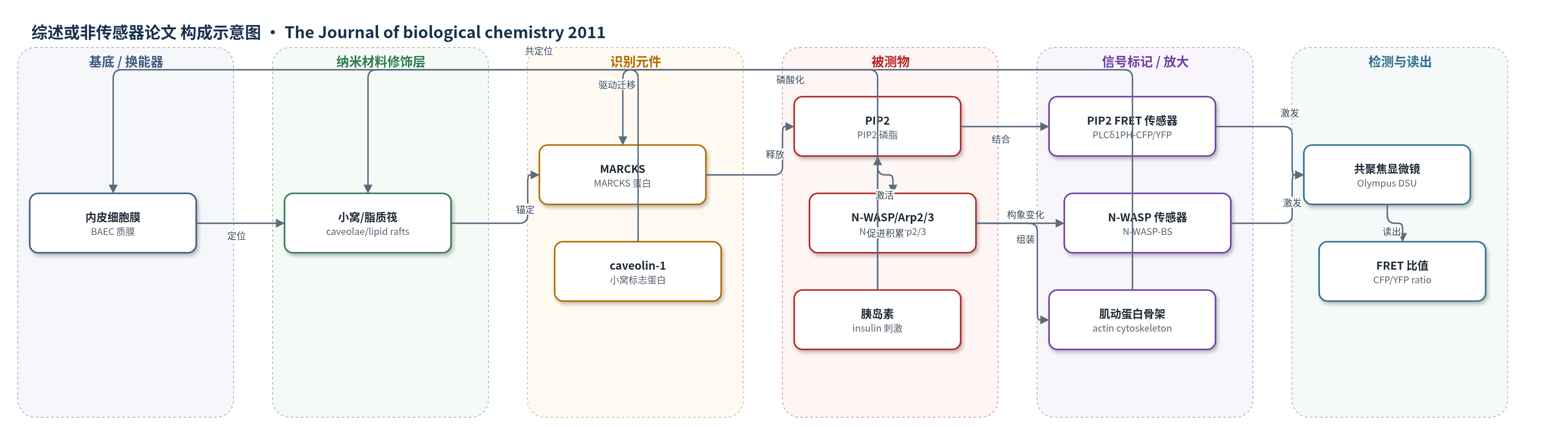
点击图片查看大图 · 依据论文自动绘制
传感器类型
综述或非传感器论文
检测对象
磷酸肌醇 4,5-二磷酸(PIP2)、N-WASP 激活状态;样品基质:培养牛主动脉内皮细胞(BAEC)活细胞、细胞裂解液及蔗糖梯度亚细胞分级。
检测原理
该文使用两种 FRET 生物传感器在活内皮细胞中监测信号事件。PIP2 传感器由 PLCδ1 PH 域分别与 CFP 和 YFP 融合组成;当 PIP2 结合 PH 域时,两个荧光蛋白靠近,CFP 激发后 YFP 发射增强,FRET 比值升高,从而反映质膜/小窝 PIP2 水平。N-WASP 传感器在 N-WASP 被激活后发生构象变化,使 FRET 比值下降,反映 N-WASP/Arp2/3 活性。胰岛素刺激内皮细胞后,MARCKS 磷酸化并从小窝转位,释放/动员 PIP2;PIP2 进一步结合并激活 N-WASP 与 Arp2/3,驱动肌动蛋白组装和定向迁移。MARCKS 敲低会削弱 PIP2 积累和 N-WASP 激活,导致 FRET 信号变化减小。
检测灵敏度
原文未报告 LOD、线性范围、灵敏度斜率或相关系数。
效应效果
研究在培养 BAEC 中验证了 MARCKS 敲低的特异性与功能效应。siRNA 敲低 MARCKS 约 90%,未改变多种内皮信号蛋白丰度;划痕愈合实验中 MARCKS 敲低显著抑制定向迁移(p<0.001,n=3),并阻断 VEGF 促进的迁移。免疫荧光显示 MARCKS 与 caveolin-1 共定位由约 70% 降至约 10%(p<0.001,n=5)。蔗糖梯度分析显示胰岛素使脂质筏/小窝 PIP2 约升高 2 倍,MARCKS 敲低削弱该升高(p<0.01,n=6)。FRET 成像显示胰岛素诱导 N-WASP 传感器 FRET 下降约 80%,MARCKS 敲低显著减弱该激活(p<0.01,n=6)。作者主张 MARCKS 是胰岛素依赖内皮 PIP2 信号与定向细胞运动的关键节点。
传感器的构成
- 样品基质:牛主动脉内皮细胞(BAEC),提供活细胞膜、小窝/脂质筏及胞内信号环境。
- 识别元件:PLCδ1 PH 域融合蛋白(PLCδ1PH-YFP 与 PLCδ1PH-CFP),特异性结合 PIP2 并作为 PIP2 传感器。
- 信号标记物:CFP/YFP 荧光蛋白对,PIP2 结合使二者靠近并产生 FRET 比值变化。
- 识别元件:N-WASP FRET 生物传感器(N-WASP-BS),通过 N-WASP 激活引起的构象变化报告活性。
- 换能/读出:Olympus DSU 共聚焦显微镜与 MetaMorph FRET 模块,CFP 激发 425 nm,检测 475 nm 与 540 nm 发射。
- 实验调控层:siRNA 敲低 MARCKS、胰岛素/VEGF 刺激及 LY294002/calphostin C 抑制,用于验证 MARCKS 依赖信号。
中文摘要
MARCKS(肉豆蔻酰化丙氨酸富集 C 激酶底物)是一种结合肌动蛋白和钙调蛋白的蛋白,广泛表达于多种哺乳动物组织,但其在内皮细胞信号反应中的作用尚不清楚。本研究在培养内皮细胞中用 siRNA 敲低 MARCKS,发现划痕愈合实验中定向细胞运动被显著抑制。作者结合生化与细胞成像方法,探讨 MARCKS 在胰岛素激活的内皮信号通路中的作用。胰岛素处理血管内皮细胞可剂量和时间依赖性地促进 MARCKS 磷酸化。细胞成像与水力学分析显示,MARCKS 定位于质膜小窝,并在胰岛素刺激下发生亚细胞转位。胰岛素还促进信号磷脂磷酸肌醇 4,5-二磷酸(PIP2)在质膜小窝中水平升高;siRNA 敲低 MARCKS 可阻断该升高,这一结果由生化检测和基于 FRET 的 PIP2 生物传感器成像共同证实。作者进一步检测 PIP2 和肌动蛋白结合蛋白 Arp2/3 与 N-WASP,发现胰岛素快速而强效地促进二者磷酸化,但 MARCKS 敲低明显削弱该反应;基于 FRET 的 N-WASP 活性生物传感器显示,MARCKS 敲低有效阻断胰岛素诱导的 N-WASP 激活。综上,MARCKS 在胰岛素依赖的内皮 PIP2 信号中起关键作用,并是血管内皮肌动蛋白组装和定向细胞运动的重要决定因素。
英文摘要
The MARCKS protein (myristoylated alanine-rich C kinase substrate) is an actin- and calmodulin-binding protein that is expressed in many mammalian tissues. The role of MARCKS in endothelial signaling responses is incompletely understood. We found that siRNA-mediated knockdown of MARCKS in cultured endothelial cells abrogated directed cell movement in a wound healing assay. We used biochemical and cell imaging approaches to explore the role of MARCKS in endothelial signal transduction pathways activated by insulin. Insulin treatment of vascular endothelial cells promoted the dose- and time-dependent phosphorylation of MARCKS. Cell imaging and hydrodynamic approaches revealed that MARCKS is targeted to plasmalemmal caveolae and undergoes subcellular translocation in response to insulin. Insulin treatment promoted an increase in levels of the signaling phospholipid phosphatidylinositol 4,5-bisphosphate (PIP(2)) in plasmalemmal caveolae. The insulin-stimulated increase in caveolar PIP(2) was blocked by siRNA-mediated knockdown of MARCKS, as determined using both biochemical assays and imaging studies using FRET-based PIP(2) biosensors. The critical role of PIP(2) in MARCKS responses was explored by examining the PIP(2)- and actin-binding proteins Arp2/3 and N-WASP. Insulin promoted the rapid and robust phosphorylation of both N-WASP and Arp2/3, but these phosphorylation responses were markedly attenuated by siRNA-mediated MARCKS knockdown. Moreover, MARCKS knockdown effectively abrogated N-WASP activation in response to insulin, as determined using a FRET-based N-WASP activity biosensor. Taken together, these studies show that MARCKS plays a key role in insulin-dependent endothelial signaling to PIP(2) and is a critical determinant of actin assembly and directed cell movement in the vascular endothelium.