组成图示
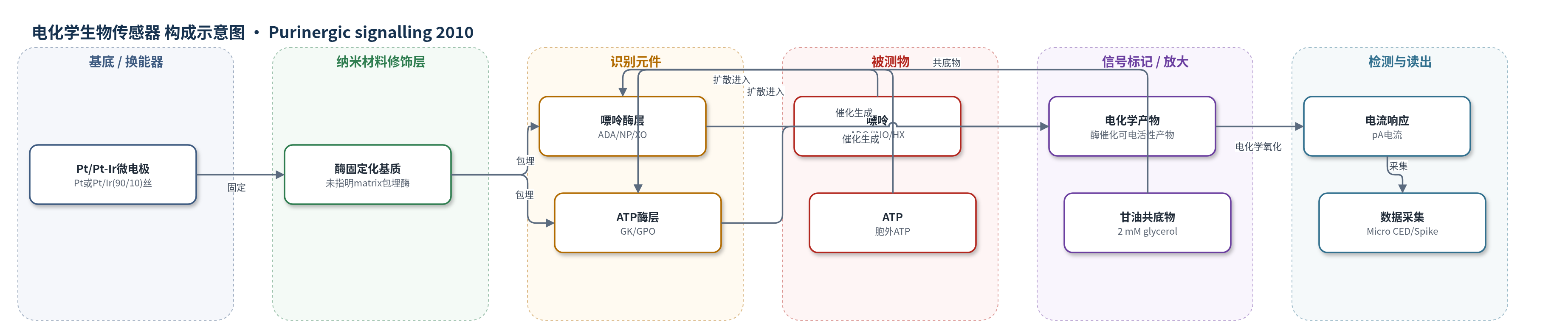
点击图片查看大图 · 依据论文自动绘制
传感器类型
电化学生物传感器
检测对象
腺苷(adenosine, ADO)、肌苷(inosine, INO)、次黄嘌呤(hypoxanthine, HX)、三磷酸腺苷(ATP);样品基质:小脑颗粒细胞原代培养物与小脑急性切片(人工脑脊液 aCSF 灌流)
检测原理
被测嘌呤(腺苷、肌苷或次黄嘌呤)从培养细胞或切片扩散至微电极表面的酶固定化基质。ADO传感器中,腺苷脱氨酶(ADA)将腺苷转化为肌苷,核苷磷酸化酶(NP)将肌苷转化为次黄嘌呤,黄嘌呤氧化酶(XO)进一步氧化次黄嘌呤,产生可在铂电极上发生氧化还原的电化学活性产物;INO传感器仅含NP和XO,主要响应肌苷/次黄嘌呤。ATP传感器利用甘油激酶(GK)与甘油-3-磷酸氧化酶(GPO)在甘油共底物存在下将ATP转化为可检测产物。酶催化产物在Pt/Pt-Ir电极表面产生法拉第电流,电流大小与局部嘌呤浓度近似线性,1 kHz采集,实现实时检测;未使用额外放大策略。
检测灵敏度
ATP检出限: ∼60 nM
效应效果
传感器经筛选后对5-HT、去甲肾上腺素、多巴胺和抗坏血酸等电活性干扰物响应显著降低;空传感器(null sensor)在刺激下几乎无电流,说明信号来自嘌呤。ADO传感器对腺苷、肌苷、次黄嘌呤灵敏度相近,响应线性且上升时间<10 s,实验前后校准可监测灵敏度漂移。25 mM KCl+1 mM谷氨酸刺激培养物时,ADO电流270±53 pA(49–813 pA),相当于1.8±0.2 μM′嘌呤,14/16盖玻片可重复;重复刺激常减少45±15%。ATP传感器未检出ATP(检出限∼60 nM)。该法首次实现原代培养细胞嘌呤释放的实时检测,并揭示释放依赖细胞组成与发育阶段。
传感器的构成
- 换能器电极:Pt或Pt/Ir(90/10)丝,蚀刻至约50 μm,作为电化学检测基底
- 酶固定化基质:未指明基质材料(matrix),包埋酶并修饰电极表面,允许嘌呤扩散
- 嘌呤识别/催化层:腺苷脱氨酶(ADA)、核苷磷酸化酶(NP)、黄嘌呤氧化酶(XO),将腺苷/肌苷/次黄嘌呤转化为可电检测产物
- 肌苷/次黄嘌呤识别/催化层:核苷磷酸化酶(NP)、黄嘌呤氧化酶(XO),响应肌苷和次黄嘌呤
- ATP识别/催化层:甘油激酶(GK)、甘油-3-磷酸氧化酶(GPO),检测ATP
- 共底物:甘油(glycerol, 2 mM),ATP检测所需共底物
- 抗干扰屏蔽:screened sensor matrix(未指明材料),降低5-HT、去甲肾上腺素、多巴胺、抗坏血酸等电活性干扰物响应
中文摘要
我们此前已报道小脑切片分子层中存在动作电位和Ca2+依赖的腺苷释放,其最可能来源是颗粒细胞轴突(平行纤维)。因此,本研究利用微电极生物传感器检测出生后第7–8天大鼠原代培养颗粒细胞能否释放腺苷。结果显示,局部电刺激不能检出嘌呤释放;但将细胞外K+浓度从3 mM升至25 mM并加入1 mM谷氨酸后,可检测到嘌呤(腺苷、肌苷或次黄嘌呤)释放。该释放机制为胞内嘌呤经均衡型核苷转运体(ENT)转运出细胞,不需要动作电位发放,但依赖Ca2+。主要释放物不是腺苷,而是肌苷或次黄嘌呤。培养物中必须同时含有颗粒细胞和胶质细胞才能发生肌苷/次黄嘌呤释放,任一细胞成分单独均不足。对出生后第7–25天小脑切片施加相同刺激也可释放嘌呤,但释放不被ENT抑制剂阻断,且发育过程中Ca2+依赖性发生改变。培养物与切片数据进一步说明嘌呤释放机制复杂,依赖细胞组成和发育阶段。
英文摘要
We have previously described an action-potential and Ca(2+)-dependent form of adenosine release in the molecular layer of cerebellar slices. The most likely source of the adenosine is the parallel fibres, the axons of granule cells. Using microelectrode biosensors, we have therefore investigated whether cultured granule cells (from postnatal day 7-8 rats) can release adenosine. Although no purine release could be detected in response to focal electrical stimulation, purine (adenosine, inosine or hypoxanthine) release occurred in response to an increase in extracellular K(+) concentration from 3 to 25 mM coupled with addition of 1 mM glutamate. The mechanism of purine release was transport from the cytoplasm via an ENT transporter. This process did not require action-potential firing but was Ca(2+)dependent. The major purine released was not adenosine, but was either inosine or hypoxanthine. In order for inosine/hypoxanthine release to occur, cultures had to contain both granule cells and glial cells; neither cellular component was sufficient alone. Using the same stimulus in cerebellar slices (postnatal day 7-25), it was possible to release purines. The release however was not blocked by ENT blockers and there was a shift in the Ca(2+) dependence during development. This data from cultures and slices further illustrates the complexities of purine release, which is dependent on cellular composition and developmental stage.