组成图示
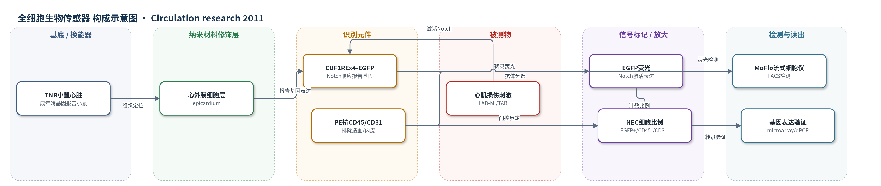
点击图片查看大图 · 依据论文自动绘制
传感器类型
全细胞生物传感器
检测对象
Notch通路转录活性(Notch pathway activity)、心肌损伤修复反应(cardiac injury repair response);样品基质:成年TNR小鼠心脏组织/心外膜细胞悬液
检测原理
Notch配体(如Delta-like 1)与受体结合后,受体胞内域入核并与CBF1/RBPJ结合,激活CBF1-REx4启动子,驱动EGFP转录。成年TNR小鼠心外膜细胞在LAD-MI或TAB等损伤刺激下Notch活性增强,EGFP蛋白累积,使EGFP+细胞数量增加。随后用PE偶联抗CD45和抗CD31抗体标记并排除造血与血管内细胞,MoFlo流式细胞仪检测EGFP荧光,计算EGFP+/CD45-/CD31-细胞占EGFP+细胞的比例(NEC%)。该比例随损伤类型和时间变化,TAB后24小时内快速升高,LAD-MI后72小时稳定并在第3-7天接近翻倍,从而将Notch通路活性转换为可定量荧光读出。
检测灵敏度
原文未报告LOD、线性范围、灵敏度斜率或R^2。
效应效果
NEC约占EGFP+细胞1%-2%,每颗对照心约10-20,000个;LAD-MI和TAB第7天NEC比例接近翻倍,TAB 24小时内快速升高,LAD-MI 72小时内稳定并于第3-7天翻倍。FACS计数与免疫组化EGFP+细胞数直接相关,作者认为可靠、有效且敏感。选择性上,NEC排除CD45+造血和CD31+内皮细胞,约50%表达Sca-1、CD105、CD44,约80%表达CD73,CD34低表达,c-Kit阴性;转录组约80%与多能基质细胞一致。功能上,EGF诱导EMT,固定化Delta-like 1激活Hes1、Hey1、Jag2、Notch1、Rbpj和Twist1,共培养或植入NOD-SCID心肌后表达α-actinin。作者主张该系统可筛选促心肌再生药物。
传感器的构成
- 报告基因层:CBF1-REx4-EGFP转基因,Notch响应元件驱动EGFP表达,报告Notch通路转录活性
- 细胞基质层:成年TNR小鼠心脏/心外膜细胞,作为活细胞传感器与损伤响应微环境
- 分选识别层:PE偶联抗CD45和抗CD31抗体,排除造血与血管内细胞,界定NEC群
- 信号标记层:EGFP荧光蛋白,Notch激活后累积,提供可定量荧光信号
- 读出装置层:MoFlo流式细胞仪(FACS),检测EGFP荧光并计算NEC比例
- 功能验证层:RetroNectin固定化Delta-like 1配体,体外激活Notch通路并诱导靶基因
中文摘要
转基因Notch报告小鼠在具有CBF1响应元件转录活性(CBF1-REx4-EGFP)的细胞中表达增强绿色荧光蛋白(EGFP),为鉴定和分离Notch激活前体细胞提供了工具。本研究询问该EGFP标记是否可定位并功能标记成年心脏组织前体细胞,以及该细胞基信号能否作为心脏损伤修复反应的定量和定性生物传感器。除散在的血管内皮和间质细胞外,Notch激活(EGFP+)细胞意外地大量富集于成年心外膜。作者用流式细胞术分离EGFP+细胞,并排除造血(CD45+)和血管内皮(CD31+)亚群,对EGFP+/CD45-/CD31-细胞进行基因表达谱和功能分析。该混合细胞池具有多能基质细胞和心外膜谱系双重特征,被命名为Notch激活心外膜来源细胞(NECs)。心肌梗死和胸主动脉缩窄扩增NEC池并促进成纤维细胞分化。克隆NEC系在血清生长因子作用下发生上皮-间质转化,固定化Notch配体Delta-like 1激活下游靶基因;与心肌细胞共培养或植入NOD-SCID小鼠心肌后,NEC心脏基因表达增加。结论:动态Notch损伤反应激活成年心外膜,产生参与纤维化修复的多能细胞群。
英文摘要
RATIONALE: Transgenic Notch reporter mice express enhanced green fluorescent protein in cells with C-promoter binding factor-1 response element transcriptional activity (CBF1-RE(x)₄-EGFP), providing a unique and powerful tool for identifying and isolating "Notch-activated" progenitors.
OBJECTIVE: We asked whether, as in other tissues of this mouse, EGFP localized and functionally tagged adult cardiac tissue progenitors, and, if so, whether this cell-based signal could serve as a quantitative and qualitative biosensor of the injury repair response of the heart.
METHODS AND RESULTS: In addition to scattered endothelial and interstitial cells, Notch-activated (EGFP(+)) cells unexpectedly richly populated the adult epicardium. We used fluorescence-activated cell sorting to isolate EGFP(+) cells and excluded hematopoietic (CD45(+)) and endothelial (CD31(+)) subsets. We analyzed EGFP(+)/CD45⁻/CD31⁻ cells, a small (<2%) but distinct subpopulation, by gene expression profiling and functional analyses. We called this mixed cell pool, which had dual multipotent stromal cell and epicardial lineage signatures, Notch-activated epicardial-derived cells (NECs). Myocardial infarction and thoracic aortic banding amplified the NEC pool, increasing fibroblast differentiation. Validating the functional vitality of clonal NEC lines, serum growth factors triggered epithelial-mesenchymal transition and the immobilized Notch ligand Delta-like 1-activated downstream target genes. Moreover, cardiomyocyte coculture and engraftment in NOD-SCID (nonobese diabetic-severe combined immunodeficiency) mouse myocardium increased cardiac gene expression in NECs.
CONCLUSIONS: A dynamic Notch injury response activates adult epicardium, producing a multipotent cell population that contributes to fibrosis repair.