组成图示
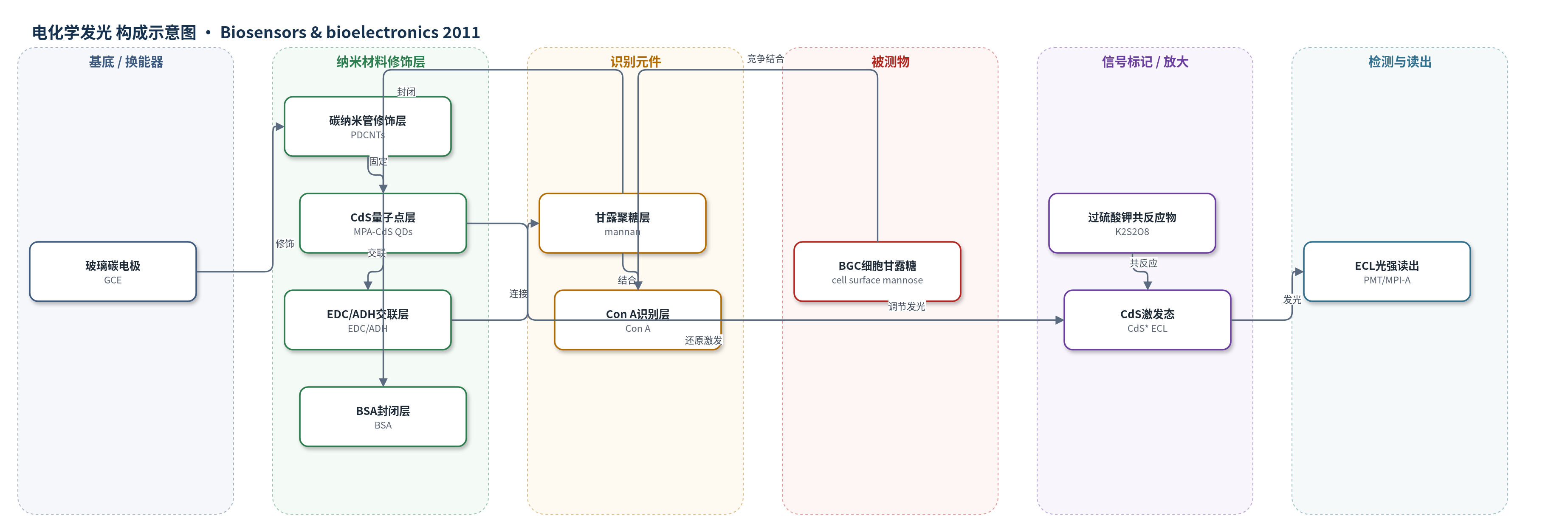
点击图片查看大图 · 依据论文自动绘制
传感器类型
电化学发光(ECL)生物传感器
检测对象
细胞表面甘露糖(cell surface mannose/mannosyl groups,以BGC细胞为模型),样品基质:PBS活细胞悬液
检测原理
电极表面甘露聚糖与刀豆蛋白A(Con A)特异性结合后形成蛋白层,阻碍CdS量子点的ECL反应,使光强降低。加入BGC细胞后,细胞表面甘露糖与电极表面甘露聚糖竞争结合Con A;细胞数量越多或表面甘露糖表达越高,电极上结合的Con A越少,ECL强度相应升高。ECL产生机制为:负电位下CdS QDs被还原为•CdS−,K2S2O8被还原生成•SO4−,二者反应向QD最高占据分子轨道注入空穴,形成激发态CdS*并发射300–650 nm光。PDCNTs提高导电性和有效面积,增强ECL信号。该无标记均相竞争策略使信号同时反映细胞量和表面碳水化合物表达水平。
检测灵敏度
LOD: 1.2 × 10^3 cells mL−1;线性范围: 2 × 10^3–1 × 10^7 cells mL−1;R = 0.997;细胞低浓度线性: 1000–8000 cells,IECL(a.u.) = 4.03 × 10^3 + 0.18n,R = 0.996;甘露糖线性: 0.1–2 pmol,IECL(a.u.) = 2.69 × 10^3 + 1.25 × 10^3 m (pmol),R = 0.998
效应效果
该传感器无需标记细胞或凝集素,可保持生物活性并避免空间位阻。对BGC细胞LOD为1.2×10^3 cells/mL,与阻抗细胞传感器检测K562细胞的1000 cells/mL相当,低于免疫芯片检测大肠杆菌O157:H7的6000 cells/mL和QCM免疫传感器检测沙门氏菌的1.0×10^4 cells/mL。重现性RSD为8.3%(1.0×10^5 cells/mL,4个电极)。4℃ PBS保存两周无明显变化,四周后保留92%初始响应。SW处理72 h后BGC细胞表面甘露糖增加33.6%,与FITC-Con A流式细胞术38.5%相符。作者认为可用于临床诊断、糖链功能研究和药物响应动态监测,并可扩展高通量检测。
传感器的构成
- 基底电极:玻璃碳电极(GCE),提供导电基底与ECL换能界面
- 导电修饰层:PDDA功能化多壁碳纳米管(PDCNTs),增强导电性、增大有效面积并固定CdS QDs
- 发光纳米材料层:巯基丙酸(MPA)封端CdS量子点(CdS QDs),作为ECL发光体
- 交联层:EDC/己二酸二酰肼(ADH),活化羧基并连接甘露聚糖
- 竞争识别层:甘露聚糖(mannan),模拟细胞表面甘露糖,与Con A特异性结合
- 封闭层:牛血清白蛋白(BSA),封闭非特异结合位点
- 识别元件:刀豆蛋白A(Con A),特异性识别甘露糖,结合后阻碍ECL
- 共反应物/电子供体:过硫酸钾(K2S2O8),还原生成硫酸根自由基,激发CdS QDs发光
中文摘要
本文设计了一种简便的电化学发光(ECL)策略,用于原位、无标记监测活细胞表面碳水化合物表达。该策略将凝集素对碳水化合物的特异性识别与碳水化合物功能化硫化镉(CdS)纳米复合材料相结合。首先将巯基丙酸(MPA)封端的CdS量子点固定在碳纳米管修饰电极上,再以甘露聚糖为模型在表面功能化碳水化合物。所得复合材料具有较高ECL灵敏度和良好稳定性,可在溶液中与目标细胞竞争结合刀豆蛋白A(Con A);由于Con A结合后阻碍ECL反应,ECL强度变化同时反映细胞数量和细胞表面碳水化合物表达水平。方法对细胞浓度在2×10^3至1×10^7 cells/mL范围内呈宽线性响应,检出限为1.2×10^3 cells/mL。该传感器可原位评价细胞表面糖链,测得单个活BGC细胞平均甘露糖基团数为8.7×10^7,并可用于监测药物处理后活细胞碳水化合物表达的动态变化。
英文摘要
A facile electrochemiluminescent (ECL) strategy for in situ label-free monitoring of carbohydrate expression on living cells was designed by integrating the specific recognition of lectin to carbohydrate with a carbohydrate-functionalized CdS nanocomposite. The mercaptopropionic acid-capped CdS quantum dots were firstly immobilized on carbon nanotubes modified electrode and then functionalized with carbohydrate using mannan as a model on the surface. The carbohydrate-functionalized CdS nanocomposite showed high ECL sensitivity and good stability, and could be used for competitive recognition to concanavalin A with the target cells in solution, which led to a change of ECL intensity due to the resistance of concanavalin A. The change depended on both the cell number and the expression level of cell surface carbohydrate. A wide linear response to cells ranging from 2×10(3) to 1×10(7) cells mL(-1) with a detection limit of 1.2×10(3) cells mL(-1) was obtained. The proposed biosensor could be used to in situ evaluate cell surface glycan, and the average number of mannose moieties on single living BGC cell was detected to be 8.7×10(7). This sensitive strategy was further used for facile monitoring of dynamic carbohydrate expression on living cells in response to drugs. The proposed method could be further expanded to high-throughput detection with the addition of more specific glycan-lectin pairs to the repertoire.