组成图示
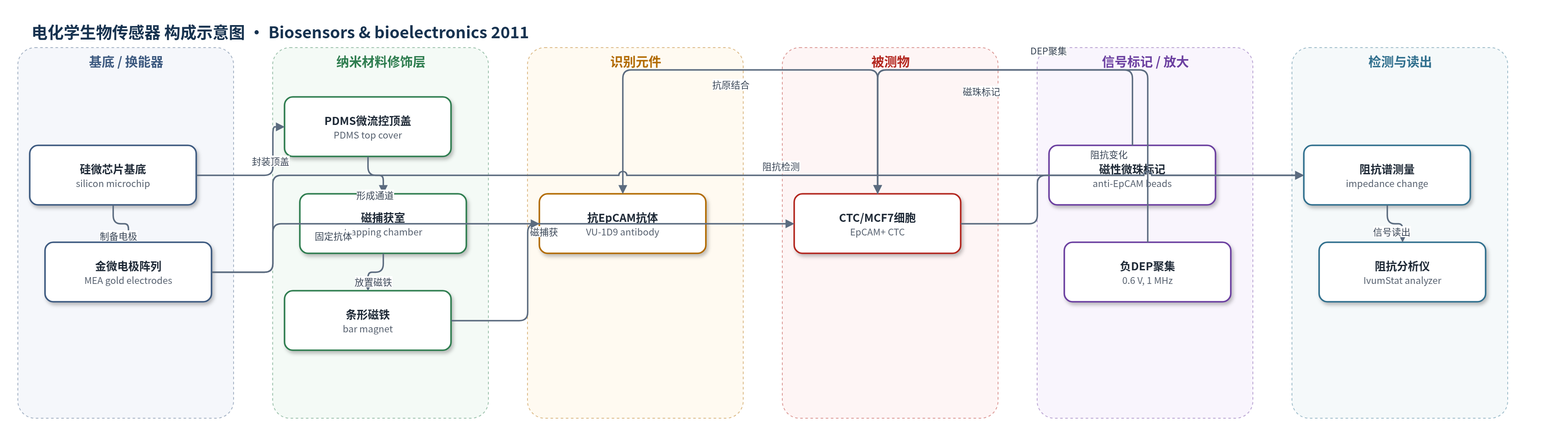
点击图片查看大图 · 依据论文自动绘制
传感器类型
电化学生物传感器
检测对象
循环肿瘤细胞(circulating tumor cells, CTC;MCF7 乳腺癌细胞);样品基质:人全血(0.5 mL 全血加标,过滤后 10 μL 血样)
检测原理
检测基于免疫磁富集与电学阻抗读出。全血先与 anti-EpCAM 磁性微珠孵育,使 CTC 表面 EpCAM 与磁珠结合;随后通过 5 μm 尺寸过滤去除红细胞、白细胞和未结合磁珠,实现 CTC 富集。富集样品进入微芯片后,背面条形磁铁在磁捕获室中捕获磁珠-CTC 复合物,血细胞被洗去;移除磁铁后复合物释放至 MEA。负 DEP(0.6 V、1 MHz)将细胞聚集到金电极中心,固定于电极的 anti-EpCAM 抗体与 CTC 表面 EpCAM 特异性结合,PBS 洗去未结合细胞和磁珠。细胞附着改变金电极-溶液界面电容与阻抗,阻抗模在约 533 kHz 出现最大变化;CTC 数量增加时,附着细胞增多,阻抗变化增大,从而通过阻抗谱区分不同 CTC 水平。
检测灵敏度
原文未报告 LOD、线性范围、灵敏度斜率或相关系数。原文报告单电极可检测低至 10 个细胞;10 μL 过滤血样中 30±2、124±29、273±23 个加标 CTC 可通过 MEA 阻抗变化区分;对应阻抗变化为 43±6、146±36、233%±52。
效应效果
系统对 0.5 mL 人全血加标 MCF7 细胞的富集回收率为 70–90%,仅加入 10 个细胞时回收率仍高于 70%;微芯片磁捕获效率平均约 90%,线性回归截距约 8 个细胞,代表过程损失阈值。三种加标水平(30±2、124±29、273±23)通过阻抗变化(43±6、146±36、233%±52)显著区分,单因素 ANOVA P<0.05。尺寸过滤、免疫磁捕获和洗涤降低了血样成分干扰,未使用荧光标记和图像分析。相比 CellSearch 等荧光法,该系统提供电子输出,适合频繁、便捷地监测治疗过程中 CTC 数量变化。
传感器的构成
- 基底/换能器电极:硅微芯片(silicon microchip)上的金微电极阵列(MEA),用于阻抗谱检测与负 DEP 聚集细胞。
- 微流控腔体:PDMS 顶盖(PDMS top cover)与芯片构成微通道、磁捕获室和 5 μL 检测室,引导样品与废液。
- 磁捕获元件:条形磁铁(bar magnet)置于芯片背面凹槽,在磁捕获室中捕获 anti-EpCAM 磁珠标记的 CTC。
- 磁性富集标记:anti-EpCAM 包被磁性微珠(magnetic beads, 3.28 μm)结合 CTC,用于免疫磁分离和芯片内磁浓缩。
- 识别元件:抗 EpCAM 抗体(anti-EpCAM antibody, VU-1D9)固定于金电极表面,特异性识别 CTC 表面 EpCAM。
- 封闭剂:BSA 封闭缓冲液(blocking buffer, BB)用于减少血样非特异结合。
- 信号读出:阻抗分析仪(IvumStat Impedance Analyzer)以 2 电极方案测量 MEA 阻抗模变化,负 DEP(0.6 V, 1 MHz)辅助细胞定位。
中文摘要
本文报道一种用于检测血液中稀有循环肿瘤细胞(CTC)的半集成电学生物传感器。样品首先通过抗 EpCAM 磁性微珠免疫磁分离与尺寸过滤进行富集,两种方法结合可获得较高富集性能,即使原始 0.5 mL 血样中仅加入 10 个 MCF7 癌细胞,回收率仍高于 70%。富集后的样品被转移至微芯片,在磁捕获室中进一步磁浓缩,随后通过固定在微电极阵列(MEA)上的抗 EpCAM 抗体进行免疫化学捕获,并利用阻抗谱进行电子检测。通过监测 MEA 阻抗变化,系统能够区分 10 μL 过滤血样中三个水平的加标 CTC 数量(30±2、124±29、273±23)。该单系统集成宏观血样处理与微尺度高灵敏电子检测,无需费时费钱的荧光标记和图像分析,有望为临床提供独立、便捷、频繁监测治疗过程中 CTC 数量变化的系统,以支持高效癌症治疗。
英文摘要
In this report, we demonstrate a semi-integrated electrical biosensor for the detection of rare circulating tumor cells (CTCs) in blood. The sample was first enriched through a combination of immunomagnetic isolation and size filtration. The integration of both methods provided a high enrichment performance with a recovery rate above 70%, even for very low numbers of cancer cells present in the original sample (10 spiked MCF7 cells in 0.5 mL of blood). In the same system, the sample was then transferred to a microchip for further magnetic concentration, followed by immunochemical trapping and electronic detection by impedance spectroscopy. Three levels of spiked CTC number (30±2, 124±29, 273±23) in 10 μL of filtered blood sample were distinguished by monitoring the impedance change of the microelectrode array (MEA). The integration of different functions in a single system provided a methodology to process milliliter-sized blood samples at the macroscale and interface with the microdimensions of a highly sensitive electronic detector. The results showed that the whole system was able to detect different levels of spiked cancer cells without the use of time- and cost-intensive fluorescence labeling and image analysis. This has the potential to provide clinicians with a standalone system to monitor changes in CTC numbers throughout therapy conveniently and frequently for efficient cancer treatments.