组成图示
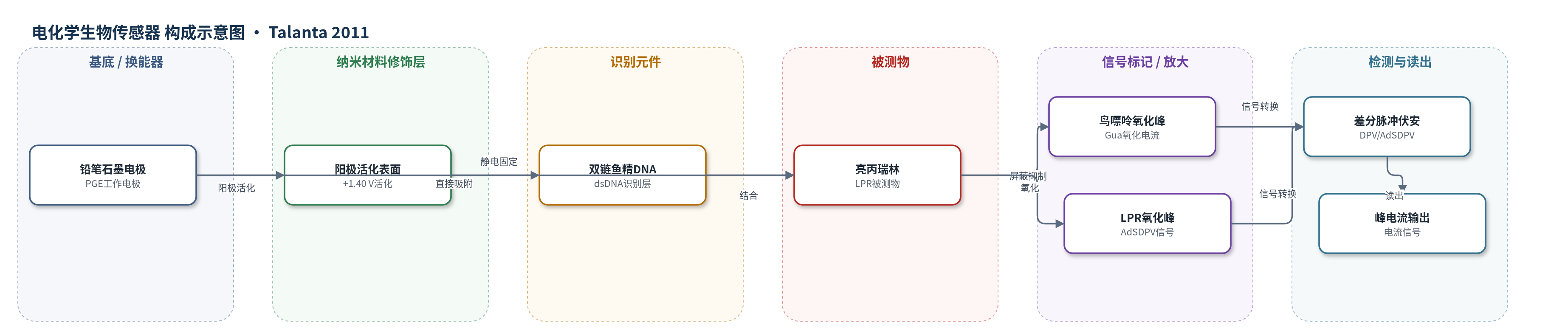
点击图片查看大图 · 依据论文自动绘制
传感器类型
电化学生物传感器
检测对象
亮丙瑞林(leuprolide, LPR);样品基质:醋酸缓冲液/BR缓冲液标准溶液及亮丙瑞林注射剂(Lucrin® injectable solution)
检测原理
该传感器以阳极活化PGE为工作电极,dsDNA通过静电作用固定于电极表面作为识别元件。LPR与dsDNA结合后,其平面芳香结构(苯酚/吲哚基团)与DNA发生相互作用,使鸟嘌呤氧化位点被屏蔽或发生损伤,导致差分脉冲伏安法(DPV)中约1.00 V处鸟嘌呤氧化峰电流下降;峰电流下降量随LPR浓度增加而线性增大。溶液相方法中,LPR先与dsDNA在溶液中结合,再于PGE上检测,同样以鸟嘌呤峰降低确认相互作用。另在裸PGE上,LPR在pH 2.00 Britton-Robinson缓冲液中经700 mV/180 s富集后发生不可逆氧化,AdSDPV剥离氧化产生峰电流,电流与LPR浓度线性相关。
检测灵敏度
裸PGE AdSDPV: LOD: 1.36 × 10^-3 ppm;线性范围: 0.005-0.20 ppm;斜率: 9.950 µA ppm^-1;相关系数: 0.997。DNA修饰PGE: LOD: 6.00 × 10^-2 ppm;线性范围: 0.20-6.00 ppm;斜率: -0.083 µA ppm^-1;相关系数: -0.987。溶液相dsDNA: LOD: 4.00 × 10^-2 ppm;线性范围: 0.20-1.00 ppm;斜率: -0.518 µA ppm^-1;相关系数: -0.995。
效应效果
该方法经全面验证,裸PGE、DNA修饰PGE和溶液相法的日内重现性RSD分别为1.55%、1.75%、1.84%,日间重现性RSD分别为2.03%、3.23%、2.52%。LPR工作标准溶液在4 ℃避光条件下可稳定至少一周,稀释溶液至少稳定10 h。三种方法直接测定Lucrin®注射剂(标示5.00 mg/mL),结果分别为4.97、5.04、4.98 mg/mL,RSD为1.89%、3.15%、2.34%,偏差为0.60%、-0.80%、0.40%;F检验和t检验表明方法间无显著差异。作者认为相比HPLC/LC-MS,该方法无需复杂样品前处理,快速、灵敏、选择性好、成本低,适合药物制剂中LPR分析。
传感器的构成
- 基底/换能器电极:一次性铅笔石墨电极(PGE),作为工作电极并传导电子。
- 电极活化层:阳极活化PGE表面(+1.40 V、60 s,0.50 M醋酸缓冲液含0.02 M NaCl,pH 4.80),形成可静电吸附DNA的表面。
- 识别元件:双链鱼精DNA(dsDNA),固定于PGE表面,与亮丙瑞林(LPR)相互作用。
- 信号指示:dsDNA中鸟嘌呤(Gua)氧化峰电流,作为LPR结合指示。
- 信号读出:差分脉冲伏安法(DPV)/吸附剥离差分脉冲伏安法(AdSDPV),输出峰电流。
中文摘要
将抗癌药亮丙瑞林(leuprolide,LPR)与固定于阳极活化铅笔石墨电极(PGE)表面的双链鱼精DNA(dsDNA)结合,用于构建一种灵敏的电化学生物传感器。采用电化学方法研究了LPR与固定于PGE上的dsDNA的相互作用,并通过差分脉冲伏安法(DPV)在PGE表面和溶液相两种相互作用方式下考察和确认其作用机制。在pH 4.80醋酸缓冲液中,以鸟嘌呤氧化峰电流的降低作为相互作用指标。对两种方法的富集时间、电位、药物浓度和重现性进行了优化。在DNA修饰PGE上,LPR浓度0.20-6.00 ppm范围内呈线性响应,检出限为0.06 ppm;溶液相相互作用法在0.20-1.00 ppm范围内线性,检出限为0.04 ppm。LPR在裸PGE上所有研究pH值下均呈不可逆氧化行为。建立了差分脉冲吸附剥离伏安法(AdSDPV)测定LPR,在0.005-0.20 ppm范围内电流与浓度线性相关,检出限为0.0014 ppm。各测定方法均经全面验证,并应用于LPR药物制剂的分析。
英文摘要
The anticancer drug, leuprolide (LPR) bound to double-stranded fish sperm DNA (dsDNA) which was immobilized onto the surface of an anodically activated pencil graphite electrode (PGE), was employed for designing a sensitive biosensor. The interaction of leuprolide (LPR) with double-stranded DNA (dsDNA) immobilized onto pencil graphite electrode (PGE) have been studied by electrochemical methods. The mechanism of the interaction was investigated and confirmed by differential pulse voltammetry using two different interaction methods; at the PGE surface and in the solution phase. The decrease in the guanine oxidation peak current was used as an indicator for the interaction in acetate buffer at pH 4.80. The response was optimized with respect to accumulation time, potential, drug concentration, and reproducibility for both interaction methods. The linear response was obtained in the range of 0.20-6.00 ppm LPR concentration with a detection limit of 0.06 ppm on DNA modified PGE and between 0.20 and 1.00 ppm concentration range with detection limit of 0.04 ppm for interaction in solution phase method. LPR showed an irreversible oxidation behavior at all investigated pH values on a bare PGE. Differential pulse adsorptive stripping (AdSDPV) voltammetric method was developed for the determination of LPR. Under these conditions, the current showed a linear dependence with concentration within a range of 0.005-0.20 ppm with a detection limit of 0.0014 ppm. Each determination method was fully validated and applied for the analysis of LPR in its pharmaceutical dosage form.