组成图示
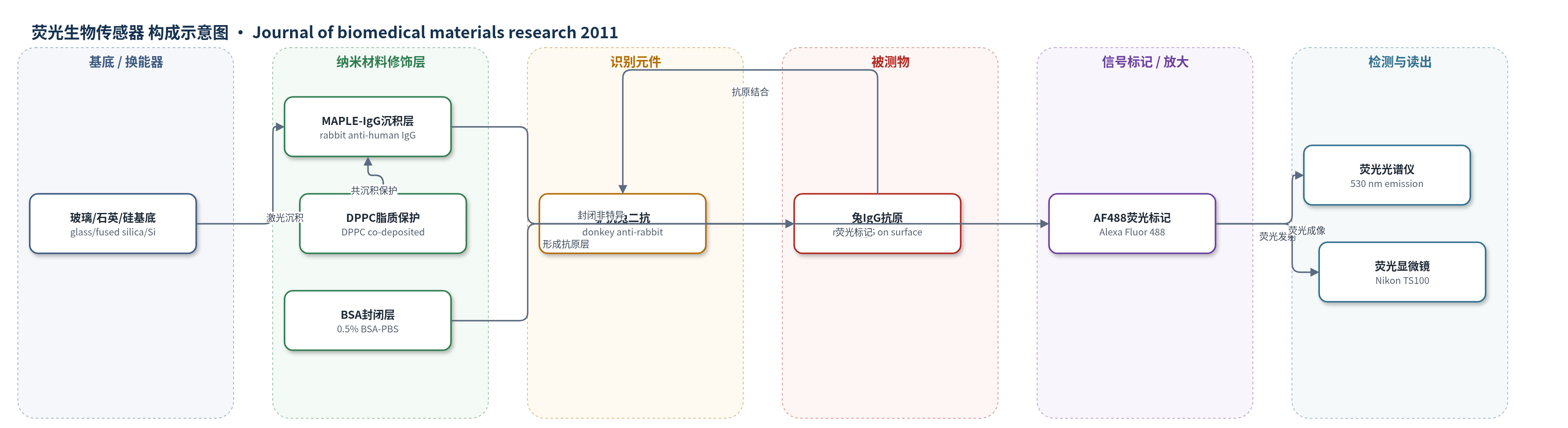
点击图片查看大图 · 依据论文自动绘制
传感器类型
荧光生物传感器
检测对象
兔免疫球蛋白G(rabbit IgG,作为抗原);样品基质:PBS/缓冲液
检测原理
本方法以玻璃、熔融石英或硅为基底,通过MAPLE将兔抗人IgG从冰冻缓冲液靶材中软转移并固定成薄膜,DPPC脂质共沉积可提高蛋白保持与界面均匀性。在验证的荧光免疫检测中,表面固定的兔IgG作为抗原,与Alexa Fluor 488标记的驴抗兔二抗特异性结合;孵育前用0.5% BSA-PBS封闭非特异位点以降低背景。AF488在激发后于530 nm发射荧光,荧光强度随表面可及兔IgG量及结合二抗量增加而增强。该体系无酶催化放大,依靠荧光标记二抗的高灵敏光学读出,通过荧光光谱或荧光显微镜成像检测信号。作者认为该固定策略可扩展为检测特定抗原的免疫传感器。
检测灵敏度
原文未报告 LOD、线性范围、灵敏度斜率或相关系数。
效应效果
选择性上,1:50 AF488驴抗兔二抗在IgG样品产生荧光点,熔融石英对照基本无信号;1:25出现非特异结合,1:200无差异。稳定性上,IgG薄膜经反复操作和液体浸没后仍固定。BCA定量显示,含脂质IL67在玻璃和熔融石英上IgG为4.10、7.52 μg,约为IL50(1.03、1.27 μg)的4倍;IL33未检出。AFM估计无脂质厚度约200、350、500 nm,含脂质约180、450、700 nm,沉积速率0.02–0.05或0.018–0.07 nm/脉冲。作者认为可用于免疫传感。
传感器的构成
- 基底/换能器:玻璃、熔融石英或硅(glass/fused silica/Si),提供支撑与表面固定
- MAPLE沉积层:兔抗人抗血清IgG(rabbit anti-human antiserum IgG),经248 nm KrF准分子激光从冰冻靶材转移形成薄膜
- 脂质修饰:L-α-磷脂酰胆碱二棕榈酰(DPPC, L-a-phosphatidylcholine dipalmitoyl),与IgG共沉积,增强固定与形态
- 固定抗原层:兔IgG(rabbit IgG),作为被荧光二抗识别的待测抗原
- 识别元件:驴抗兔二抗(donkey anti-rabbit secondary antibody),特异性结合兔IgG
- 信号标记物:Alexa Fluor 488(AF488),共轭于驴抗兔二抗,产生荧光
- 封闭剂:0.5%牛血清白蛋白-PBS(BSA-PBS),封闭非特异结合位点
- 缓冲/清洗:PBS/生理缓冲液(TRIS 50 mM, pH 7.4, NaCl 150 mM),用于孵育、清洗与检测
中文摘要
本文报道了利用基质辅助脉冲激光蒸发(MAPLE)将兔抗人抗血清免疫球蛋白G(IgG)进行保护性转移并固定。将含0.2–2 mg/mL IgG、是否添加脂质(L-α-磷脂酰胆碱二棕榈酰,DPPC)的蒸馏水/生理缓冲液冰冻成靶材,在室温下用准分子激光辐照,使玻璃、熔融石英或硅基底上形成薄IgG涂层。每个样品施加10000次0.33、0.5或0.67 J/cm²激光脉冲。采用光学显微镜、扫描电镜、原子力显微镜和傅里叶变换红外光谱研究薄膜形貌与组成。由于荧光方法对低量抗原(IgG)灵敏,选用荧光光谱和荧光显微镜验证生物传感器换能原理。样品浸入驴抗兔二抗溶液后,证明所有结构均实现蛋白固定。在MAPLE溶液中加入脂质可改善IgG的转移与固定。
英文摘要
The sheltered transfer and immobilization of rabbit anti-human antiserum immunoglobulin G (IgG) by matrix-assisted pulsed laser evaporation (MAPLE) are reported. The iced targets submitted to laser irradiation consisted of 0.2-2 mg/mL IgG blended or not with lipid (L-α-phosphatidylcholine dipalmitoyl) dissolved in distilled water-based saline buffer. Thin IgG coatings were obtained at room temperature onto glass, fused silica, or silicon substrates. Ten thousand subsequent laser pulses of 0.33, 0.5, or 0.67 J/cm(2) fluence were applied for the synthesis of each sample. Morphology and composition of the thin films were studied by optical, scanning, and atomic force microscopy and Fourier transformed infrared spectrometry. Optical labeling methods such as spectrofluorimetry and fluorescence microscopy were selected to verify the biosensor transduction principle because of their high sensitivity for detecting low amounts of antigen (IgG). Protein immobilization to the substrate surface was demonstrated for all obtained structures after immersion in the donkey anti-rabbit secondary antibody solution. The IgG transfer and immobilization onto substrates were improved by addition of lipid to MAPLE solutions.