组成图示
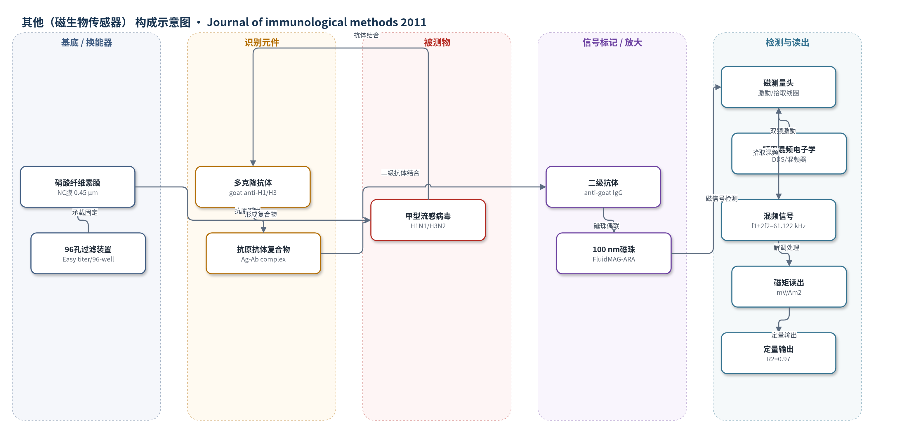
点击图片查看大图 · 依据论文自动绘制
传感器类型
其他(磁生物传感器)
检测对象
甲型流感病毒H1N1(Influenza A H1N1, A/Beijing/262/95)、甲型流感病毒H3N2(Influenza A H3N2, A/Kiev/301-94);样品基质:PBS稀释的灭活病毒抗原/96孔微孔板
检测原理
病毒抗原先被硝酸纤维素膜吸附,随后加入H1N1或H3N2特异性多克隆抗体,在膜内形成抗原–抗体复合物;再加入偶联兔抗山羊IgG的100 nm磁珠,使膜上结合的磁珠数量与病毒量成正比。磁传感器采用双频激励:f1=61 kHz、f2=61 Hz,磁珠在低磁场下具有强非线性磁化,产生f1+2f2=61,122 Hz混频分量;差分拾取线圈选择性检测该分量,而顺磁/抗磁材料的线性响应被抑制。最终输出电压随磁珠/病毒浓度升高而增大,实现无酶定量检测。
检测灵敏度
LOD: 3.12 μg/200 μl(磁珠);线性范围: 20.0–4.0 μg/200 μl(磁珠);R^2 = 0.97;病毒校准范围: 210.0 pg/200 μl(H1N1)、450.0 pg/200 μl(H3N2);R^2 = 0.97(H1N1)、0.98(H3N2);噪声等效磁矩: 1.0×10⁻¹³ Am2/√Hz
效应效果
新方法与常规ELIFA和FITC荧光点印迹法(DBA)相比,检测能力无显著差异,可检测每孔皮克级病毒,但省去显色和酸终止两步,缩短检测时间并提高稳健性。空白信号相对标准偏差方面,磁珠法H1N1为1.67%、H3N2为1.20%,低于ELIFA的4.98%和1.69%;DBA为1.83%和1.38%。磁珠法标准差为H1N1 9.45 mV、H3N2 7.07 mV,ELIFA为0.08 OD和0.02 OD,DBA为723 RFU和552 RFU。校准曲线R^2分别为0.97/0.98(磁珠法)、0.90/0.96(ELIFA)、0.97/0.99(DBA)。作者认为该方法可兼容96孔批量样品,样品测后可保存,适合现场快速、稳定、可重复的免疫检测,但膜非特异结合和磁珠堵塞孔隙仍需优化。
传感器的构成
- 固相载体:硝酸纤维素膜(nitrocellulose membrane,孔径0.45 μm),用于吸附病毒抗原并提供较大内表面
- 识别元件:H1N1/H3N2多克隆抗体(goat polyclonal antibody),与膜上病毒抗原特异性结合
- 信号标记物:兔抗山羊IgG(Fc-specific secondary antibody)偶联100 nm磁珠(FluidMAG-ARA),作为磁性标记
- 封闭剂:含1.0% BSA封闭缓冲液(blocking buffer),降低非特异结合
- 换能器:磁测量头(magnetic measurement head),含两个激励线圈和一个差分拾取线圈,检测磁珠非线性磁化
- 读出电子学:DDS芯片(AD9834)、混频器、放大器、滤波器和A/D,检测f1+2f2=61,122 Hz混频分量
中文摘要
本文报道了一种基于硝酸纤维素膜和频率混频磁生物传感器检测两种甲型流感病毒的新分析方法。该方法先将病毒抗原吸附于硝酸纤维素膜上,再加入特异性多克隆抗体形成抗原–抗体复合物,随后用偶联二级抗体的磁珠进行标记,最后通过磁传感器检测膜上结合的磁珠。与常规ELISA/ELIFA相比,该方法省去显色和终止反应两个步骤,可缩短检测时间并提高方法稳健性。作者将新方法与常规ELIFA及FITC荧光点印迹法进行比较,结果表明在优化条件下可检测每孔皮克级病毒,适用于快速、稳定、可重复的现场检测。
英文摘要
Here we describe a new analytical method for the detection of two influenza A viruses by nitrocellulose membrane and magnetic sensors that employ a special frequency mixing technique. The combination of the nitrocellulose membrane and magnetic bead detection permits a rapid assay procedure and excludes two steps (the development of color and the stop reaction) required for usual immunochemical detection methods such as ELISA. Quantitative virus detection was performed using magnetic beads conjugated with secondary antibody. The results were compared with conventional assay methods and with a dot-blot assay with fluorescence compound (FITC). Under optimum conditions, our new assay procedure is capable of detecting picograms of virus per well. This new method combining the nitrocellulose membrane and magnetic bead detection reduces analytical time and allows stable and repeatable analyses of samples in point-of-care applications.