组成图示
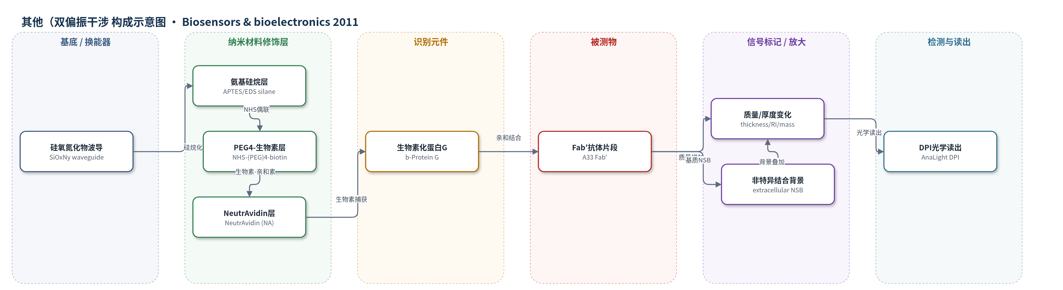
点击图片查看大图 · 依据论文自动绘制
传感器类型
其他(双偏振干涉(DPI)光学生物传感器)
检测对象
Fab′片段抗体(A33 Fab′ / fragment antibody Fab′);样品基质:大肠杆菌补料分批发酵液/胞外培养基(fermentation broth / extracellular medium),纯化Fab′在PBS-T中用于校准
检测原理
该传感器采用双偏振干涉(DPI)无标记光学检测。硅氧氮化物波导经氨基硅烷化后,用NHS-(PEG)4-biotin共价引入生物素,再捕获NeutrAvidin(NA),最后捕获生物素化蛋白G(b-ProG)。当样品中的Fab′与b-ProG亲和结合时,传感表面生物层质量、厚度和折射率增加;632.8 nm激光探测波导中双偏振光相位变化,DPI将相位转换为厚度、折射率和单位面积质量。Fab′浓度越高,结合量越大,DPI响应越大,经四参数剂量响应曲线定量。表面可用pH 2.5磷酸盐再生。复杂发酵液需4倍稀释以降低基质干扰。
检测灵敏度
LOD: 1.7 µg/ml;分析范围: 10–1000 µg/ml;动态范围: 两个数量级
效应效果
传感器在PBS-T中对纯化Fab′的分析范围为10–1000 µg/ml,LOD为1.7 µg/ml。21次注入/再生循环中,100 µg/ml Fab′响应为0.69±0.03 rad,再生后0.0013±0.0002 rad,约100%再生。4倍稀释发酵液加标回收率为82.4–89.1%,2倍稀释为53.7–65.3%,未稀释为43.5–51.9%,说明需稀释降低基质干扰。诱导后2 h即可检测Fab′泄漏,阈值0.020 rad;30 h内表面可再生,之后出现污染。单次分析约2 min,快于HPLC约7 min。监测显示20 h后泄漏加速,Fab′从60 mg/l升至294 mg/l,提示收获点。
传感器的构成
- 基底/换能器:硅氧氮化物玻璃波导(SiOxNy waveguide,FB 80),作为DPI光学换能器,探测表面层厚度与折射率变化
- 表面清洁/活化:酸洗或UV/臭氧处理,去除有机污染并暴露羟基,便于硅烷化
- 氨基硅烷修饰层:3-aminopropyltriethoxysilane(APTES)与N-[3-(trimethoxysilyl)propyl]ethylenediamine混合硅烷,共价结合到波导表面并提供氨基
- 生物素连接层:NHS-(PEG)4-biotin(优选)或sulfo-NHS-LC-biotin,经NHS与氨基偶联,提供生物素位点;PEG亲水链改善表面有序性
- 亲和捕获层:NeutrAvidin(NA),通过生物素-亲和素结合非共价固定,提供生物素结合位点并降低非特异结合
- 识别元件:biotinylated Protein G(b-ProG),被NA捕获,特异性识别Fab′抗体片段
- 被测物/样品:A33 Fab′片段抗体,来自大肠杆菌发酵液/胞外培养基,纯化Fab′用于校准
- 再生/清洗:PBS-T运行缓冲液和20 mM磷酸氢二钠(pH 2.5)再生液,洗脱Fab′并恢复传感表面
- 信号读出:DPI仪器(AnaLight)测量相位、厚度、折射率和质量变化,转换为Fab′浓度
中文摘要
本文报道了一种基于双偏振干涉(DPI)的多层生物传感器表面,用于监测大肠杆菌补料分批发酵中抗体片段(Fab′)泄漏至胞外培养基后的浓度。该传感器以生物素化蛋白G(b-ProG)为识别层,通过NeutrAvidin(NA)和生物素连接层固定在硅氧氮化物波导上。作者利用DPI原位分析各传感层的活性、稳定性和功能,并用X射线光电子能谱(XPS)和二次离子质谱(SIMS)离线评估化学层的组成与均一性。两种生物素连接物产生显著不同的表面:亲水NHS-(PEG)4-biotin使b-ProG层有序沉积在NA层上方,形成活性且稳定的表面;疏水sulfo-NHS-LC-biotin使b-ProG层埋入NA层,导致表面不均一且Fab′结合较差。该传感器检测限为1.7 µg/ml,动态范围覆盖两个数量级,可在诱导后2 h检测到Fab′泄漏,信噪比高且胞外成分干扰小。Fab′泄漏呈双相,诱导后20 h转为更快速率,提示产品损失加速并需及时收获。
英文摘要
This paper describes the step-wise fabrication and characterisation of a multi-layer dual polarization interferometry (DPI) based biosensor utilising Protein G (ProG) as the bio-recognition layer for the detection of a fragment antibody (Fab'). The biosensor is capable of monitoring the concentration of Fab' product within the extracellular medium of a fed-batch fermentation after leakage from Escherichia coli (E.coli). The activity, stability and functionality of each sensor layer were analysed in situ using DPI, whilst the chemical identity and homogeneity of the chemical layers were assessed ex situ using X-ray photoelectron spectroscopy (XPS) and secondary ion mass spectrometry (SIMS). Two different biotin linkers were found to produce hugely differing surfaces after the capture of NeutrAvidin™ (NA) and biotinylated Protein G (b-ProG). The hydrophilic (PEG)(4)-biotin linker resulted in a surface where the b-ProG layer was deposited and organised above the NA layer producing an active and stable surface, whilst the hydrophobic LC-biotin linker generated a surface where the b-ProG layer was buried within the NA layer leading to variable surfaces and poor binding of the Fab' target. The biosensor has a detection limit of 1.7 μg/ml with a dynamic range covering two orders of magnitude. The sensor can detect the onset of Fab' leakage as early as 2h following product induction, with high signal-to-noise ratios and little interference from extracellular components. Leakage of Fab' followed a biphasic profile, switching to a more rapid rate 20 h after induction, indicating accelerated product loss and the need for cultivation harvest.